Search
Invitrogen
Arginase 1 Monoclonal Antibody (A1exF5), PerCP-eFluor™ 710, eBioscience™
{{$productOrderCtrl.translations['antibody.pdp.commerceCard.promotion.promotions']}}
{{$productOrderCtrl.translations['antibody.pdp.commerceCard.promotion.viewpromo']}}
{{$productOrderCtrl.translations['antibody.pdp.commerceCard.promotion.promocode']}}: {{promo.promoCode}} {{promo.promoTitle}} {{promo.promoDescription}}. {{$productOrderCtrl.translations['antibody.pdp.commerceCard.promotion.learnmore']}}
Additional Information:
{{banner.description}}
")
FIGURE: 1 / 24
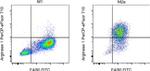
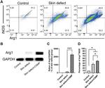
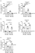
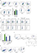
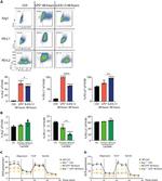
Arginase 1 Antibody (46-3697-82) in Flow
C57BL/6 mouse bone marrow derived macrophages were polarized for 24 hours with either LPS (Product # 00-4976-93) and IFN-gamma (Product # 16-7311-85) to make M1 macrophages (left) or with IL-4 (Product # BMS338) to make M2a macrophages (right). Cells were surface stained with F4/80 Monoclonal Antibody, FITC (Product # 11-4801-82) then stained intracellularly, using the Intracellular Fixation & Permeabilization Buffer Set (Product # 88-8824-00) and protocol, with 0.125 µg Arginase 1 Monoclonal Antibody, PerCP-eFluor 710. Total viable cells ... View More

Please note: We are reviewing Western blot images included in the antibody testing data in our catalog, including those provided by third parties. Unless expressly labeled or annotated as “raw-unedited”, Western blot images included in the antibody testing data in our catalog may have been edited, optimized or otherwise adjusted for presentation.
 in Flow")
 in IHC")
 in Flow")
 in Flow")
 in Flow")
 in Flow")
 in Flow")
 in Flow")
 in Flow")
 in Flow")
 in Flow")
 in Flow")
 in Flow")
 in Flow")
 in Flow")
 in Flow")
 in Flow")
 in Flow")
 in Flow")
 in Flow")
 in Flow")
 in Flow")
 in Flow")
 in Flow")
Product Details
46-3697-82
Applications
Tested Dilution
Publications
Product Specifications
Species Reactivity
Human,
Mouse
Published species
Human,
Mouse
Host/Isotype
Rat
/ IgG2a, kappa
Recommended Isotype Control
Class
Monoclonal
Type
Antibody
Clone
A1exF5
Immunogen
E.coli-derived Recombinant mouse Arginase 1
Conjugate
PerCP-eFluor™ 710
PerCP-eFluor™ 710
PerCP-eFluor™ 710
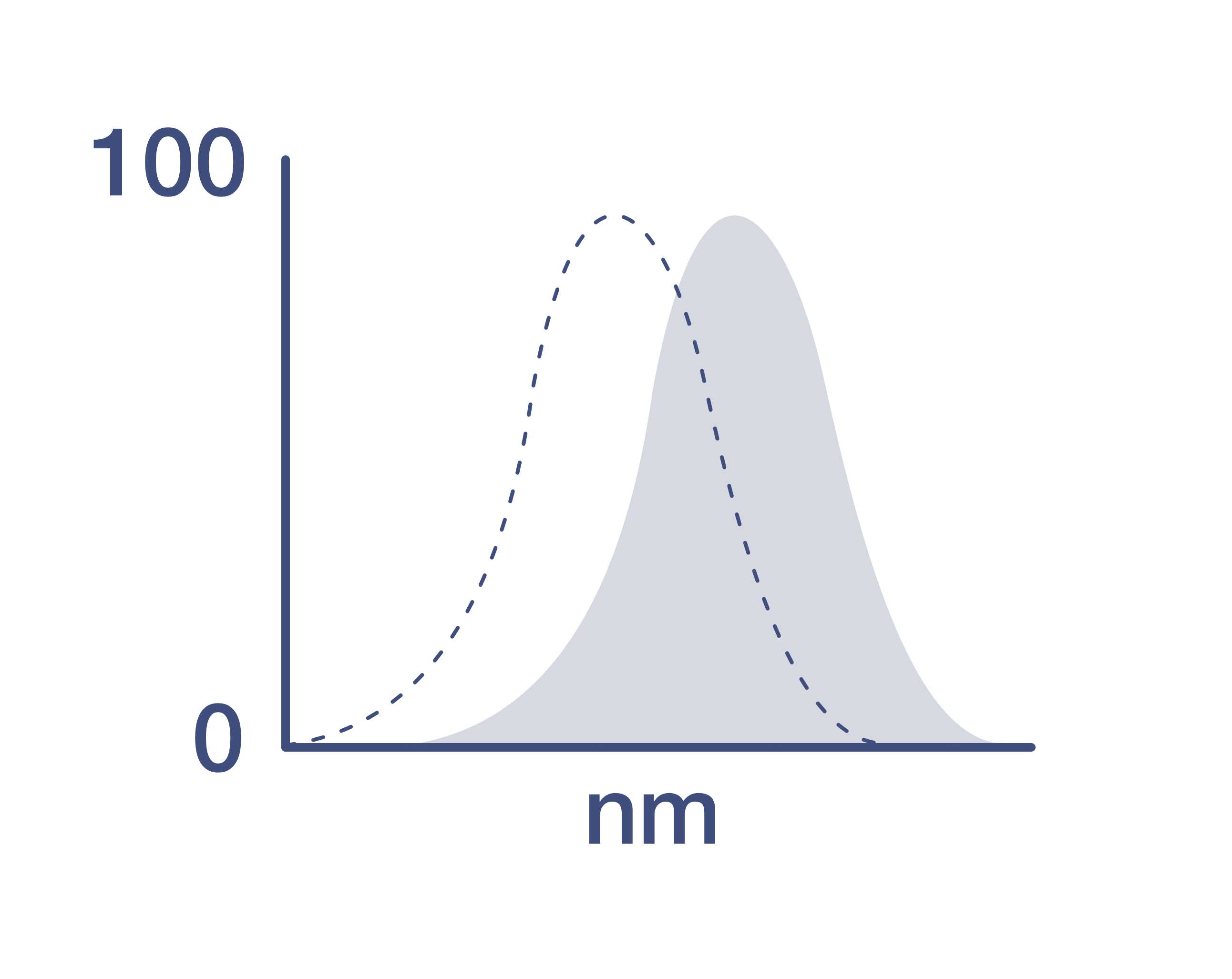
Excitation/Emission Max
482/708 nm
View spectra
Form
Liquid
Concentration
0.2 mg/mL
Amount
100 µg
Purification
Affinity chromatography
Storage buffer
PBS, pH 7.2
Contains
0.09% sodium azide
Storage conditions
4°C, store in dark, DO NOT FREEZE!
Shipping conditions
Ambient (domestic); Wet ice (international)
RRID
Product Specific Information
Description: The monoclonal antibody A1exF5 recognizes both human and mouse Arginase 1, a cytosolic enzyme (Arg1). This A1exF5 clone is compatible with both, the standard intracellular protocols, and the Foxp3/Transcription Factor Staining Buffer Set.
Applications Reported: This A1exF5 antibody has been reported for use in flow cytometric analysis.
Applications Tested: This A1exF5 antibody has been tested by flow cytometric analysis of stimulated mouse bone marrow cells using the Intracellular Fixation & Permeabilization Buffer Set (Product # 88-8824-00) and protocol. Please refer to BestProtocols®: Protocol A: Two step protocol for (cytoplasmic) intracellular proteins located under the Resources Tab online. This may be used at less than or equal to 0.25 µg per test. A test is defined as the amount (µg) of antibody that will stain a cell sample in a final volume of 100 µL. Cell number should be determined empirically but can range from 10^5 to 10^8 cells/test. It is recommended that the antibody be carefully titrated for optimal performance in the assay of interest.
PerCP-eFluor™ 710 emits at 710 nm and is excited with the blue laser (488 nm); it can be used in place of PerCP-Cyanine5.5. We recommend using a 710/50 bandpass filter, however, the 695/40 bandpass filter is an acceptable alternative. Please make sure that your instrument is capable of detecting this fluorochrome.
Light sensitivity: This tandem dye is sensitive to photo-induced oxidation. Please protect this vial and stained samples from light.
Fixation: Samples can be stored in IC Fixation Buffer (Product # 00-8222) (100 µL of cell sample + 100 µL of IC Fixation Buffer) or 1-step Fix/Lyse Solution (Product # 00-5333) for up to 3 days in the dark at 4°C with minimal impact on brightness and FRET efficiency/compensation. Some generalizations regarding fluorophore performance after fixation can be made, but clone specific performance should be determined empirically.
Excitation: 488 nm; Emission: 710 nm; Laser: Blue Laser.
Target Information
Arginase-1 (Arg1) is a 35 kDa enzyme converting L-arginine to urea and L-ornithine, which is the final step in the urea cycle. The resulting polyamines are important for cell proliferation and removal of toxins that arise from protein degradation. By degrading arginine, Arginase 1 deprives NO synthase of its substrate and down-regulates nitric oxide production. In both human and mouse, Arginase 1 is expressed in the liver, neutrophils, myeloid derived suppressor cells (MDSC) and neural stem cells. In human, expression in blood neutrophils but not in CCR3+ granulocytes has been reported. In mice, expression of Arginase 1 is one of the hallmarks of alternatively activated macrophages (M2a). Arginase-1 may be expressed in the myeloid cells infiltrating tumors, and is typically found in the majority of hepatocellular carcinomas. Defects in Arginase 1 are the cause of argininemia, an autosomal recessive disorder characterized by hyperammonemia.
For Research Use Only. Not for use in diagnostic procedures. Not for resale without express authorization.
How to use the Panel Builder
Watch the video to learn how to use the Invitrogen Flow Cytometry Panel Builder to build your next flow cytometry panel in 5 easy steps.
Bioinformatics
Protein Aliases: A-I; Arginase; arginase 1 liver; arginase 1, liver; arginase I; Arginase-1; Arginase1; HGNC:663; Liver Arginase; Liver-type arginase; Type 1 Arginase; Type I arginase
Gene Aliases: AI; AI256583; Arg-1; ARG1; PGIF
UniProt ID: (Mouse) Q61176
Entrez Gene ID: (Mouse) 11846
Disclaimer

Clicking the images or links will redirect you to a website hosted by BenchSci that provides third-party scientific content. Neither the content nor the BenchSci technology and processes for selection have been evaluated by us; we are providing them as-is and without warranty of any kind, including for use or application of the Thermo Fisher Scientific products presented.

Performance Guarantee
If an Invitrogen™ antibody doesn't perform as described on our website or datasheet,we'll replace the product at no cost to you, or provide you with a credit for a future purchase.*
Learn more
We're here to help
Get expert recommendations for common problems or connect directly with an on staff expert for technical assistance related to applications, equipment and general product use.
Contact tech support