Search
Invitrogen
DST Polyclonal Antibody
{{$productOrderCtrl.translations['antibody.pdp.commerceCard.promotion.promotions']}}
{{$productOrderCtrl.translations['antibody.pdp.commerceCard.promotion.promocode']}}: {{promo.promoCode}} {{promo.promoTitle}} {{promo.promoDescription}}. {{$productOrderCtrl.translations['antibody.pdp.commerceCard.promotion.learnmore']}}
{{$ctrl.translations['antibody.pdp.skuNotes.additionalInformation']}}:
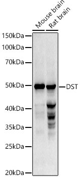
FIGURE: 1 / 1
DST Antibody (PA5-96034) in WB
Western blot analysis of DST in extracts of various cell lines. Samples were incubated with DST Polyclonal antibody (Product # PA5-96034) using a dilution of 1:1,000, followed by HRP Goat Anti-Rabbit IgG (H+L) at a dilution of 1:10,000. Lysates/proteins: 25 µg per lane. Blocking buffer: 3% nonfat dry milk in TBST. Detection: ECL Basic Kit. Exposure time: 90s.
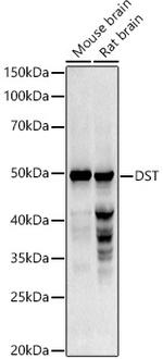
Please note: We are reviewing Western blot images included in the antibody testing data in our catalog, including those provided by third parties. Unless expressly labeled or annotated as “raw-unedited”, Western blot images included in the antibody testing data in our catalog may have been edited, optimized or otherwise adjusted for presentation.

Product Details
PA5-96034
Product Specifications
Species Reactivity
Human,
Mouse,
Rat
Host/Isotype
Rabbit
/ IgG
Class
Polyclonal
Type
Antibody
Immunogen
Recombinant protein (or fragment)
Conjugate
Unconjugated
Unconjugated
Unconjugated
Form
Liquid
Concentration
1.98 mg/mL
Amount
198 µg
Purification
Affinity Chromatography
Storage buffer
PBS, pH 7.3, with 50% glycerol
Contains
0.09% sodium azide
Storage conditions
-20°C, Avoid Freeze/Thaw Cycles
Shipping conditions
Ambient (domestic); Wet ice (international)
RRID
Product Specific Information
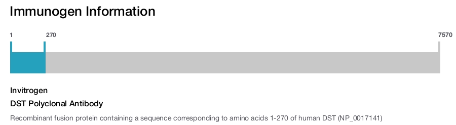
Immunogen sequence: MHSSSYSYRS SDSVFSNTTS TRTSLDSNEN LLLVHCGPTL INSCISFGSE SFDGHRLEML QQIANRVQRD SVICEDKLIL AGNALQSDSK RLESGVQFQN EAEIAGYILE CENLLRQHVI DVQILIDGKY YQADQLVQRV AKLRDEIMAL RNECSSVYSK GRILTTEQTK LMISGITQSL NSGFAQTLHP SLTSGLTQSL TPSLTSSSMT SGLSSGMTSR LTPSVTPAYT PGFPSGLVPN FSSGVEPNSL QTLKLMQIRK PLLKSSLLDQ; Positive Samples: THP-1, Rat kidney; Cellular Location: Cell junction, Cell membrane, Cell projection, Cytoplasm, Endoplasmic reticulum membrane, H zone, Lipid-anchor, Membrane, Nucleus, Nucleus envelope, Single-pass membrane protein, Z line, axon, cell cortex, cytoskeleton, cytoskeleton, hemidesmosome, myofibril, sarcomere
Target Information
As basal cells of stratified squamous epithelia begin to migrate in response to wound healing, they lose their cell-substrate adhesion junctions, the hemidesmosomes. The hemidesmosome is an adhesion structure of the epidermal-dermal junction in keratinocytes. When keratinocytes migrate laterally or upward to differentiate they must control the formation and disintegration of the hemidesmosomes. The bullous pemphigoid antigen BPAG1 is a hemidesmosomal protein of the cutaneous basement membrane zone. The primary sequence deduced from full-length human cDNAs predicts that this molecule consists of a central rod region and flanking globular domains. A neuronal isoform, BPAG1n3 is the result of differential splicing of BPAG1. BPAG1n3 is distinguished by its initial 32 amino acid residues and by the absence of the amino-terminal half of the actin-binding domain.
For Research Use Only. Not for use in diagnostic procedures. Not for resale without express authorization.
References (0)
Have you cited this product in a publication?
Let us know so we can reference it here.
Bioinformatics
Protein Aliases: BPA; BPAG1a3; BPAG1eA; MACF-like product; BPAG1eB; second IF-binding domain product; bullous pemphigoid antigen; bullous pemphigoid antigen 1; cytoskeletal protein; cytoskeletal protein BPAG1; dystonia musculorum protein; hemidesmosomal plaque protein; microtubule actin cross-linking factor 2; trabeculin-beta; unnamed protein product
Gene Aliases: 2310001O04Rik; A830042E19Rik; ah; athetoid; BP230; BP240; BPA; Bpag; BPAG1; BPAG1-n; CATX-15; CATX15; CMYO29; D6S1101; DMH; DT; EBS3; EBSB2; HSAN6; LCCS12; MACF2; mKIAA0728; nmf203; nmf339
UniProt ID: (Human) Q03001, (Mouse) Q91ZU6
Entrez Gene ID: (Human) 667, (Mouse) 13518, (Rat) 316313
microtubule cytoskeleton organization
cytoskeleton organization
axonogenesis
retrograde axonal transport
response to wounding
maintenance of cell polarity
regulation of microtubule polymerization or depolymerization
cytoplasmic microtubule organization
hemidesmosome assembly
wound healing
intermediate filament cytoskeleton organization
intracellular transport
cell motility
integrin-mediated signaling pathway
Disclaimer

Clicking the images or links will redirect you to a website hosted by BenchSci that provides third-party scientific content. Neither the content nor the BenchSci technology and processes for selection have been evaluated by us; we are providing them as-is and without warranty of any kind, including for use or application of the Thermo Fisher Scientific products presented.

Performance Guarantee
If an Invitrogen™ antibody doesn't perform as described on our website or datasheet,we'll replace the product at no cost to you, or provide you with a credit for a future purchase.*
Learn more
We're here to help
Get expert recommendations for common problems or connect directly with an on staff expert for technical assistance related to applications, equipment and general product use.
Contact tech support