Global impacts of the chikungunya virus
Chikungunya is a viral illness spread by mosquitoes in Africa, Asia, and the Americas, with occasional cases appearing in different areas. The disease induces fever and intense joint pain, which can be crippling, and lasts for varying periods of time. Dengue and Zika share similar symptoms with chikungunya, leading to frequent misdiagnoses.
The Alphavirus chikungunya virus (CHIKV) is the pathogen responsible for this severe illness. CHIKV forms unique organelles known as spherules on the infected cell’s plasma membrane to facilitate viral RNA replication. While the structures of several viral proteins within the spherules have been identified, the complete structure of the organelle remains unknown.
Cryo-electron microscopy of virus structure
Researchers at Umea University utilized cryo-electron tomography to investigate the structural organization of the spherules, combining the high-resolution imaging of a Thermo Scientific Krios Cryo-TEM (cryo-transmission electron microscope) with Thermo Scientific Amira Software for 3D visualization, segmentation, and analysis. The 3D reconstruction showed that there were clusters of balloon-shaped spherules attached to the plasma membrane.

Figure 1. 3D reconstruction of CHIKV spherules at the plasma membrane. Yellow: plasma membrane, red: viral RNA, blue: protein complexes found at the spherule necks. Figure reproduced from Laurent et al. under CC BY 4.0.
The circular spherules were found to be 50 to 70 nm in diameter. The tomograms also revealed a filament structure within the membrane buds. The position and dimensions of these filaments indicate that they are likely viral RNA, potentially in its double-stranded replicative form.
Understanding the structure of viral filaments in CHIKV spherules
The study sought to determine if observation of the visible filaments within the spherules could help estimate the amount of viral RNA in each organelle. Automated filament tracing in Amira Software was used to trace and quantify the filaments in the individual spherules. This proved to be an effective way to both quantify and analyze the structure of the organelles.

Figure 2. Characterization and quantification of viral RNA in CHIKV spherules. A) 3D structure of the dsRNA filaments obtained by Amira Software, where yellow is the membrane, red is RNA, and blue is the neck complex base. B) Increase of RNA length relative to spherule volume. C) Estimation of dsRNA length (in base pairs) for two datasets, along with the average number of copies per spherule. Figure reproduced from Laurent et al. under CC BY 4.0.
The total length of viral filaments within the spherules was found to be 18,600±2,900 Å and 21,400±1,600 Å per spherule (Figure 2B), which is equivalent to about 7,300±1,150 and 8,400±600 base pairs per spherule (Figure 2C). This length corresponds to roughly 80-90% of a full-length replicon RNA (8,820 base pairs), suggesting that each spherule likely contains one complete copy of the template RNA strand, with a significant portion in a double-stranded form. This is a pivotal observation for deconvoluting the replication mechanisms of the virus and could inform future research and therapeutic strategies.
The study of these RNA filaments offers detailed insights into their organization within the spherules. The authors also explored protein and lipid interactions, revealing that non-structural proteins (nsP1 – nsP4) play a significant role in stabilizing the spherule structure and facilitating RNA replication. Characterization of lipid composition, meanwhile, highlighted the importance of specific lipids in membrane curvature and stability. These findings reveal the interplay between RNA, proteins, and lipids in maintaining the structural integrity and functionality of viral replication organelles.
Thermo Scientific microscopes and software reveal virus structure
Thermo Scientific Amira Software provides advanced visualization, segmentation, and analytical capabilities for the examination and interpretation of intricate datasets, such as those produced with cryo-electron tomography.
Laurent et al. demonstrated how Amira Software enables the 3D visualization of virus structures by analyzing Chikungunya spherules at the plasma membrane. Further quantitative analyses even allowed them to estimate the viral RNA content of the spherules.
Amira Software is designed to manage large datasets and generate accurate 3D reconstructions along with quantitative analysis, greatly enhancing research with precise and comprehensive results.
Learn more at thermofisher.com/amira
References
- World Health Organization Chikungunya Fact Sheet. URL: https://www.who.int/news-room/fact-sheets/detail/chikungunya
- Laurent, T, Architecture of the chikungunya virus replication organelle.eLife 11:e83042 (2022). doi: 10.7554/eLife.83042

Advances in high-resolution cryo-EM at 100 kV
100 kV cryo-TEM enables high-resolution single particle anal... Alex Ilitchev, PhD
Read More
3D Tissue Histology with Light-Sheet Microscopy Enables Nondestructive Analysis of Microglia
3D tissue analysis offers critical benefits for neuroscience... Alex Ilitchev, PhD
Read More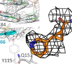
Fragment based drug discovery meets challenging drug targets with high-throughput cryo-EM
Benefits of FBDD in the search for novel therapeutics Frag... Dominic Meusch
Read More
Targeted protein degradation as a novel therapeutic approach for undruggable diseases
Induced proximity for targeted protein degradation In 1993, ... Dominic Meusch
Read More
Leave a Reply