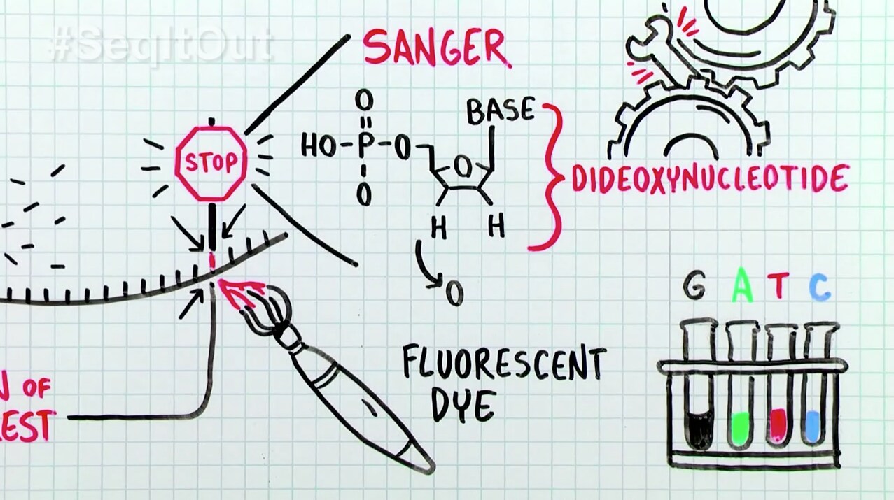
Let’s go back to the basics and explore the technology platform that has been regarded as the gold standard for many years. You guessed it – we’re talking about Sanger Sequencing by capillary electrophoresis. Many might ask, “why is it called Sanger Sequencing?” Sanger Sequencing is named after the inventor of this ground breaking technology, Dr. Frederick Sanger, who developed this method over 40 years ago in the mid-70s. So, what are the basics of Sanger Sequencing?
It all starts by having a short primer binding next to the region of interest. In the presence of the 4 nucleotides, the polymerase will extend the primer by adding on the complementary nucleotide from the template DNA strand. To find the exact composition of the DNA sequence, we need to bring this reaction to a defined stop that allows us to identify the base of the very end of this particular DNA fragment. Sanger did this by removing an oxygen atom from the ribonucleotide. Such a nucleotide is called a dideoxynucleotide. This is analogous to throwing a wrench into a gear. The polymerase enzyme can no longer add normal nucleotides onto this DNA chain. The extension has stopped and we now need to identify what it is. We identify the chain terminating nucleotide by a specific fluorescent dye, 4 specific colors to be exact. Sanger sequencing results in the formation of extension products of various lengths terminated with dideoxynucleotides at the 3′ end.
The extension products are then separated by Capillary Electrophoresis or CE. The molecules are injected by an electrical current into a long glass capillary filled with a gel polymer. During CE, an electrical field is applied so that the negatively charged DNA fragments move toward the positive electrode. The speed at which a DNA fragment migrates through the medium is inversely proportional to its molecular weight. This process can separate the extension products by size at a resolution of one base. A laser excites the dye labeled DNA fragments as they pass through a tiny window at the end of the capillary. The excited dye emits a light at a characteristic wavelength that is detected by a light sensor. Software can then interpret the detected signal and translate it into a base call. When the sequencing reaction is performed in the presence of all four terminated nucleotides, you eventually get a pool of DNA fragments that are measured and separated base by base. What you will get in the end is a data file showing the sequence of the DNA in a colorful electropherogram and a text file which you can use to answer the questions you may be asking.
I’m a junior in college studying biology and this was the most understanding of Sanger sequencing I’ve had since learning about it freshman year. Thanks so much! Sending this to my friends :)


Your content with steps is one of the best I’ve read! Thank you for that!
I’m a junior in college studying biology and this was the most understanding of Sanger sequencing I’ve had since learning about it freshman year. Thanks so much! Sending this to my friends :)
Why is the beginning of a sequencing read messy? And why does it run out by ~1000 bp?
Wow ! Awesome video. Thank you !
잘 읽어 습니다..
새로운 것을 알게 되어 감사 합니다.