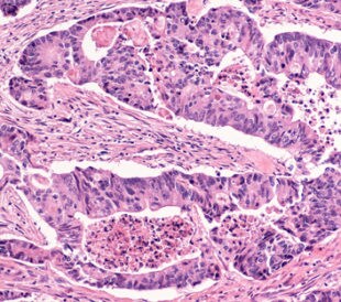
One of the attractive features of proteomics is the opportunity to identify disease biomarkers. Unfortunately, the field has been hindered by the necessity of extracting small amounts of information from relatively large sample sizes and the consequent necessity of analyzing protein changes in only a few proteins at a time. In terms of adenocarcinoma and colorectal cancer, proteomics once had limited application in determining alterations in signaling pathways, the cell-surface proteome, and the way genetic information is processed in tumor cells.
Wisniewski et al.1 assert methodological advances that address these issues. Typical proteomics methods have required a large sample, limiting the opportunity for researchers to use microdissection techiniques. Innovative approaches allowed Wisniewski et al. to identify and compare membrane proteins derived from a sample of only a few micrograms of protein. Additionally, the researchers used high-accuracy mass spectrometry (Thermo Scientific) to analyze the proteomes and posttranslational modifications of FFPE samples of normal, cancerous, and metastasized mucosal tissues. In this way, the researchers evaluated the subcellular components, molecular function, and signaling pathways at play in CRC and demonstrated that the proteome experiences a high degree of change between normal (N) and cancerous (C) cells, yet experiences very little change between becoming cancerous and metastatic (M).
Analyzing archival, microdissected FFPE tissues, the researchers identified over 8,000 proteins, 99% of which were found in the N, C, and M stages of CRC samples. Of these proteins, the researchers found that, accounting for variations based on the relative abundance of each protein, the proteome experiences a 50% change between normal tissues and cancerous tissues. In particular, normal cells contain 20% to 30% more extracellular and plasma membrane proteins than do adenocarcinoma cells, which experience a 50% decrease in channel proteins and a 30% decrease in transporter proteins. Normal cells also contain 30% fewer nuclear proteins since cancer cells demonstrate a 2- to 10-fold increase in transcription factors and chromatin activators. Interestingly, the researchers found no significant difference in cytoplasmic proteins.
In terms of biomarkers, researchers used unsupervised clustering to identify 1,808 proteins that were upregulated or downregulated in cancer cells. During this process, the normal cells clustered together while the cancerous and metastatic cells clustered in patient-specific pairs. This indicates that tumor cells are more similar to their own metastasized twin than they are to other tumor cells. The upregulated proteins, especially the 34 upregulated plasma membrane proteins the researchers found, may prove to be excellent biomarkers for CRC. Wisniewski et al. state that future research should include the investigation of these upregulated plasma membrane proteins as secreted proteins in plasma samples since they could then be observed over time using traditional techniques, such as ELISA.
Wisniewski et al. quantified specific proteins that were changed in the signaling pathways of tumor cells. They found that 95% of the altered proteins that participate in regulating transcription and translation were upregulated. Cancer cells are known for increased glycolysis, and the researchers identified 11 altered proteins involved in that process. They also identified an 80% downregulation of altered proteins that influence oxidative phosphorylation. In general, the researchers found that extracellular proteins and transporter or channel proteins as well as plasma membrane channel proteins were all downregulated in adenocarcinoma cells, while plasma membrane receptor proteins were upregulated.
The researchers also used cultured cells in order to compare these more reproducible results to those found in the microdissected tissues. Using a method they call total protein approach, or TPA, Wisniewski et al. estimated the absolute copy numbers for the proteins in the enterocytes. To do this, the researchers compared individual LFQ intensities to the total MS signal and then divided these values by the molecular weight and multiplied them by both the Avogadro constant and the protein content of a single cell. The copy numbers obtained using this method were comparable to those derived from more traditional methods, indicating that TPA is a reliable method for estimating copy numbers from diverse data sets. On both clinical and laboratory levels, these innovative approaches to proteomics represent valuable assets in the evaluation and quantification of proteome remodeling in tumor cells, as well as the search for novel biomarkers for CRC.
References
1. Wisniewski, J., et al. (2012) ‘Extensive quantitative remodeling of the proteome between normal colon tissue and adenocarcinoma‘, Molecular Systems Biology, 8 (611), (pp. 1-15)
New Diagnostic Biomarkers in Colorectal Cancer
Colonoscopies are currently the best method to diagnose colo...
Read More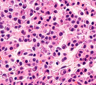
Top-Down Proteomic Characterization of Histone H3 Proteoforms in Disease
Zheng et al. performed top-down mass spectrometric proteomic...
Read More
Effects of Rosemary Extract on Colon Cancer Cells
A team of researchers from Spain and Sweden recently ...
Read More
Lung Cancer Biomarker Discovery through Metabolic Enzyme Activity
Sun et al. (2016) present a biomarker discovery study that s...
Read More
Leave a Reply