 Angi et al. (2016) present a nano-liquid chromatography–tandem mass spectrometry (nanoLC-MS/MS) approach to biomarker discovery for uveal melanoma (UM) diagnosis.1 Using label-free quantitation, the research team characterized tumor secretomes released into conditioned media by UM primary cell culture, and then filtered the results to indicate potentials as circulating biomarkers.
UM, the most common intraocular tumor in adults, carries a very guarded prognosis. Diagnosis is difficult, and 50% of cases develop metastatic spread to the liver that is invariably fatal. Treatment options are limited. Since pathogenesis often involves proteins secreted by the tumor itself that either propagate survival or influence normal tissues for invasion, Angi et al. propose that biomarker discovery among cancer cell products is a valid approach.
The researchers obtained UM tumors from patients diagnosed with the disease. Based on chromosome 3 characteristics, four of these showed low metastatic potential, whereas 10 posed a high risk of spread. The team compared results obtained from short-term primary cell cultures of these tumors with preparations established using normal choroidal melanocytes (NCM) obtained from five healthy donors. The study thus comprised three sample groups: UM-HR (high metastatic risk), UM-LR (low metastatic risk) and NCM.
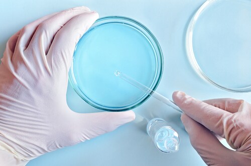
Once established, Angi et al. confirmed cell type by characterizing the morphology of the cultures in situ. They then collected conditioned media from all cultures using standard methods, harvesting the secretome by taking the supernatant following centrifugation to separate the cell bodies and debris. The team enriched and concentrated the low-abundance proteins in the conditioned media with a bead adsorption step. Following on-bead digestion they analyzed the peptides by nanoLC-MS/MS using an UltiMate 3000 RSLCnano system coupled with a Q Exactive mass spectrometer (both Thermo Scientific) operating in data-dependent acquisition mode.
Overall, the team identified 1,843 proteins across all three sample cultures. With further data filtering, followed by functional analysis, they were able to classify 539 of these as secreted. Protein profiles were similar across all three sample types. However, cluster analysis showed a strong demarcation between samples for all but one UM-HR culture. The researchers note that this culture was the only one to undergo passaging before analysis.
The researchers applied more stringent filtering to the MS data, sorting according to a false detection rate of 1% and excluding proteins identified by fewer than three unique peptides. They then re-examined the results for differential expression, quantifying proteins by comparing average individual abundance with results across sample runs and between sample groups. This sorting process resulted in a list of 325 proteins with differential abundance when compared with the NCM cultures. Of these, 163 proteins overlapped between UM-HR and UM-LR cultures, with 83 increased in abundance and 80 showing reduced levels. Examination of the data showed that most proteins with increased levels in UM-conditioned media were either classically or non-classically secreted.
When Angi et al. examined the data from the HR-UM cells, they found 53 proteins showing significantly altered levels. Of these, 33 proteins showed upregulation and 20 were decreased. Functional and Ingenuity Pathway Analysis for enrichment terms showed that 32 proteins derived from exosomes (15 with increased abundance levels; 17 with reduced levels). Most of these were involved in extracellular matrix remodeling, cell migration and invasion. The team determined that 13 of these proteins showed potential for further evaluation as circulating biomarkers for UM.
Reference
1. Angi, M., et al. (2016) “In-depth proteomic profiling of the uveal melanoma secretome,” Oncotarget [Epub ahead of print], doi: 10.18632/oncotarget.10418.

New Diagnostic Biomarkers in Colorectal Cancer
Colonoscopies are currently the best method to diagnose colo...
Read More
Top-Down Proteomic Characterization of Histone H3 Proteoforms in Disease
Zheng et al. performed top-down mass spectrometric proteomic...
Read More
Effects of Rosemary Extract on Colon Cancer Cells
A team of researchers from Spain and Sweden recently ...
Read More
Lung Cancer Biomarker Discovery through Metabolic Enzyme Activity
Sun et al. (2016) present a biomarker discovery study that s...
Read More
Leave a Reply