The ability to identify and quantify protein isoforms in a complex mixture is a growing challenge in proteomics research. Protein isoforms can arise through a mutation in the coding region, resulting in an amino acid substitution, deletion, or insertion of additional amino acids; alternative splicing of messenger RNA; a co- or posttranslational maturation event, such as proteolytic cleavage of a leader peptide; or posttranslational modifications that affect the structure or function of a protein, such as phosphorylation. At every step in a protein’s life cycle, a modification, error, or degradation can occur that can affect that proteins localization, function, or half-life.
Processing of messenger RNA through maturation or alternative splicing leads to a mixture of transcripts for the ribosome to translate. This heterogeneous mixture of RNA is what gives rise to many of the protein isoforms present in eukaryotic organisms. Some proteins have several isoforms that arise through this mechanism. Tau, a protein involved in neurodegenerative disorders, exists in six distinct isoforms. Furthermore, these six isoforms are posttranslationally modified through a variety of mechanisms, including phosphorylation.1 Hyperphosphorylation of different isoforms of the Tau protein could be indicative of Alzheimer’s disease or several different neurodegenerative disorders. Understanding which phosphorylation states or which isoforms are present in these various diseases, as well as the ability to accurately detect and quantify these forms, can help in the development of reliable markers of the neurodegenerative process for diagnosis and treatment.
One standard way in proteomics research to measure the presence or number of protein isoforms in a cell is to perform two-dimensional gel electrophoresis. In two-dimensional gel electrophoresis, proteins are separated by isoelectric point and then by mass. If a protein is modified by phosphorylation, there will be several spots that correspond to the same protein because the phosphate group can change the isoelectric point. Also, if there are isoforms present due to alternative splicing, they will also have unique molecular weights and isoelectric points.2,3 In Tran et al., an improvement over the standard two-dimensional gel electrophoresis procedure on the separation and isolation of proteins is described. The procedure uses two-dimensional liquid electrophoresis in the liquid fraction of intact proteins by isoelectric point followed by gel-eluted liquid fraction and entrapment for fractionation by molecular weight. This fractionation is followed by additional reverse-phase nanocapillary liquid chromatography and mass spectrometry with an LTQ Fourier Transform Ultra Mass Spectrometer (Thermo Scientific).3
Using this “four-dimensional” platform, the most comprehensive top-down proteomic survey was achieved on HeLa cells. Almost 3,100 protein isoforms / species were detected using this technique. The nearly 3,100 protein identifications came from 1,043 individual proteins. Several posttranslational modifications were also identified including 645 phosphorylations, 538 lysine acetylations, 158 protein methylations, 19 lipid attachments, and 5 hypusines. This technique sidesteps some of the inherent shortcomings of the two-dimensional electrophoresis approach to isoform separation and identification: mainly, the amount of sample required. This technique requires less sample due to smaller scale isolation and greater recovery of the initial sample.3
Continuation of growth in proteomics research requires faithful and accurate assignment of a complex mixture of proteins and protein isoforms. Tran et al. demonstrate that this platform can yield amazingly high resolution data on a mixture of intact proteins as complex as a nuclear or cytosolic fraction from a cancer cell line. Increased knowledge of number and posttranslational state of different isoforms and the ability to measure the covariance in modification patterns in different tissues or disease states will aid in the understanding of intracellular signaling and increase the effectiveness of biomarker screening and drug development. Understanding protein isoforms and posttranslational modification will become a cornerstone of proteomics research in the years to come.
References
1. Buée, L., et al. (2000) ‘Tau protein isoforms, phosphorylation and role in neurodegenerative disorders‘, Brain Research Reviews, 33 (1), (pp. 95-130)
2. Blakeley, P., et al. (2010) ‘Investigating protein isoforms via proteomics: a feasibility study‘, Proteomics, 10 (60), (pp. 1127-1140)
3. Tran, J.C., et al. (2011) ‘Mapping intact protein isoforms in discovery mode using top-down proteomics‘, Nature, 480 (7376), (pp. 254-258)
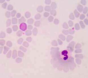
Acetylation of the Malaria Parasite
Lysine acetylation is a ubiquitous post-translational modifi...
Read More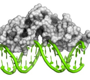
Holocaust Exposure and Intergenerational Impact via FKBP5 Methylation
Scientists know that, in animal models, stress can produce e...
Read More
C-Terminomics: What’s Happening at the End of the Rainbow
Structurally speaking, all proteins have a beginning and an ...
Read More
New Roles for Lysine Demethylase KDM4A
Lysine methyltransferases and lysine demethylases are two ex...
Read More
Thanks for the nice description and references, this is very useful for my students.