Search
Invitrogen
Human IL-33 Recombinant Protein
{{$productOrderCtrl.translations['antibody.pdp.commerceCard.promotion.promotions']}}
{{$productOrderCtrl.translations['antibody.pdp.commerceCard.promotion.viewpromo']}}
{{$productOrderCtrl.translations['antibody.pdp.commerceCard.promotion.promocode']}}: {{promo.promoCode}} {{promo.promoTitle}} {{promo.promoDescription}}. {{$productOrderCtrl.translations['antibody.pdp.commerceCard.promotion.learnmore']}}
Additional Information:
{{banner.description}}

")
产品信息
RP-8623
产品规格
Expression System
E. coli
分类
Recombinant
类型
Protein
偶联物
Unconjugated
Unconjugated
Unconjugated
形式
Lyophilized
浓度
0.1 mg/mL
Amount
10 µg
保存条件
-20°C, Avoid Freeze/Thaw Cycles
运输条件
Wet ice
产品详细信息
Recombinant human IL-33 is a non-glycosylated protein, containing 160 amino acids, with a molecular weight of 18.1 kDa. Purity is typically greater than 95% and endotoxin level less than 1 EU/ug.
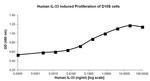
The expected biological activity (ED50) is determined by the ability to induce the proliferation of D10S cells and is typically less than 0.1 ng/mL. The specific activity of this protein is 1x10^7 units/mg.
Reconstitute using sterile water at 0.1 mg/mL and centrifuge prior to opening vial. Gently pipet solution down the sides of the vial. DO NOT VORTEX sample. Store reconstituted material at -20°C and add 0.1% BSA for additional stability.
Amino acid sequence: MSITGISPIT EYLASLSTYN DQSITFALED ESYEIYVEDL KKDEKKDKVL LSYYESQHPS NESGDGVDGK MLMVTLSPTK DFWLHANNKE HSVELHKCEK PLPDQAFFVL HNMHSNCVSF ECKTDPGVFI GVKDNHLALI KVDSSENLCT ENILFKLSET.
靶标信息
IL-33 (Interleukin-33) is a 270 amino acid, highly divergent protein belonging to the IL-1 family with an IL-1-like C-terminal domain. IL-33 is a dual function protein that may function both as a proinflammatory cytokine and an intracellular nuclear factor with transcriptional regulatory properties. IL-33 binds to and signals through IL1RL1/ST2 and its stimulation recruits MYD88, IRAK1, IRAK4, and TRAF6. IL-33 activates NF-kappaB and MAP kinases, and drives production of TH2-associated cytokines from in vitro polarized TH2 cells. In vivo, IL-33 induces the expression of IL-4, IL-5, and IL-13 and leads to severe pathological changes in mucosal organs. IL-33 is proteolytically converted to a mature form by CASP1 and is highly expressed in high endothelial venules found in tonsils, Peyer's patches and mesenteric lymph nodes and is almost undetectable in placenta. Prolonged IL-33 treatment of mice led to the development of eosinophilia, splenomegaly, and severe pathological changes in mucosal organs such as lungs, esophagus and small intestine. Recent experiments have shown that IL-33 can also co-localize with heterochromatin and possesses transcriptional repressor activities, indicating that IL-33 may function as both a proinflammatory cytokine and an intracellular nuclear factor with transcriptional regulatory properties. Despite its predicted molecular weight, IL-33 will often run at higher molecular weight in SDS-PAGE. Studies have shown that IL-33 can also co-localize with heterochromatin and possesses transcriptional repressor activities, indicating that IL-33 may function as both a proinflammatory cytokine, and an intracellular nuclear factor with transcriptional regulatory properties.
仅用于科研。不用于诊断过程。未经明确授权不得转售。
篇参考文献 (0)
您是否在文献中引用过该产品?请点击下方按钮邮件告知我们。
Disclaimer

Clicking the images or links will redirect you to a website hosted by BenchSci that provides third-party scientific content. Neither the content nor the BenchSci technology and processes for selection have been evaluated by us; we are providing them as-is and without warranty of any kind, including for use or application of the Thermo Fisher Scientific products presented.
