Search


引用和文献 (6)
Gibco™
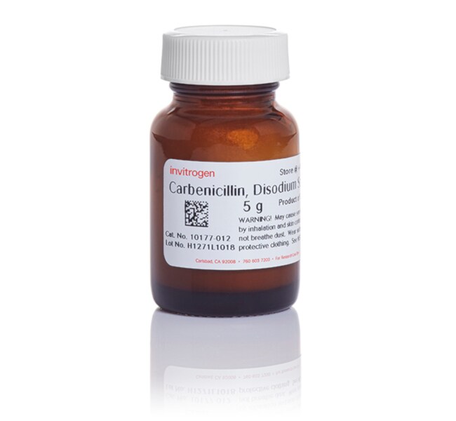
羧苄青霉素二钠盐
羧苄青霉素二钠盐是一种具有羧基和苯甲基的半合成青霉素抗生素。其作用机制与氨苄青霉素相同。干扰革兰氏阴性细菌的细胞壁合成,同时对植物组织显示出低毒性。Gibco 羧苄青霉素用作耐药性农杆菌和大肠杆菌的选择性抗生素,通常浓度为 50–100 µg/mL。β-内酰胺酶 (bla) 基因对氨苄青霉素具有耐药性,也对羧苄青霉素具有耐药性了解更多信息
| 货号 | 数量 |
|---|---|
| 10177012 | 5 g |
货号 10177012
价格(CNY)
7,203.00
Each
数量:
5 g
价格(CNY)
7,203.00
Each
羧苄青霉素二钠盐是一种具有羧基和苯甲基的半合成青霉素抗生素。其作用机制与氨苄青霉素相同。干扰革兰氏阴性细菌的细胞壁合成,同时对植物组织显示出低毒性。Gibco 羧苄青霉素用作耐药性农杆菌和大肠杆菌的选择性抗生素,通常浓度为 50–100 µg/mL。β-内酰胺酶 (bla) 基因对氨苄青霉素具有耐药性,也对羧苄青霉素具有耐药性。与氨苄青霉素相比,β-内酰胺酶对其分解更慢,因此更稳定。在长期孵育期间可减少卫星菌落的生长。本产品以粉末形式提供,应制成 50–100 mg/mL 水储备液。
仅供科研使用。不可用于诊断程序。
规格
颜色白色、类白色
最大浓度50-100 μg/mL
产品线Gibco
数量5 g
有效期36 个月
运输条件湿冰
形式粉末
产品类型Geneticin
无菌无菌
Unit SizeEach
内容与储存
储存条件:2 至 8°C
运输条件:冰
有效期:自生产之日起 36 个月
运输条件:冰
有效期:自生产之日起 36 个月
常见问题解答 (FAQ)
我该如何对我的培养物去污染?
你们提供哪些抗生素来帮助用户控制或减少细胞培养中的污染情况?
What are the recommended concentrations of antibiotics to use for selection in prokaryotes and eukaryotes?
Can ampicillin be used for selection of eukaryotic cells if put under control of a eukaryotic/viral promoter?
How can I decontaminate my cultures?
引用和文献 (6)
引用和文献
Abstract
The RNA Helicase DDX6 Controls Cellular Plasticity by Modulating P-Body Homeostasis.
Journal:Cell Stem Cell
PubMed ID:31588046
'Post-transcriptional mechanisms have the potential to influence complex changes in gene expression, yet their role in cell fate transitions remains largely unexplored. Here, we show that suppression of the RNA helicase DDX6 endows human and mouse primed embryonic stem cells (ESCs) with a differentiation-resistant, "hyper-pluripotent" state, which readily reprograms to
Nudt21 Controls Cell Fate by Connecting Alternative Polyadenylation to Chromatin Signaling.
Journal:Cell
PubMed ID:29249356
'Cell fate transitions involve rapid gene expression changes and global chromatin remodeling, yet the underlying regulatory pathways remain incompletely understood. Here, we identified the RNA-processing factor Nudt21 as a novel regulator of cell fate change using transcription-factor-induced reprogramming as a screening assay. Suppression of Nudt21 enhanced the generation of induced
High-throughput, image-based screening of pooled genetic-variant libraries.
Journal:Nat Methods
PubMed ID:29083401
We report a high-throughput screening method that allows diverse genotypes and corresponding phenotypes to be imaged in individual cells. We achieve genotyping by introducing barcoded genetic variants into cells as pooled libraries and reading the barcodes out using massively multiplexed fluorescence in situ hybridization. To demonstrate the power of image-based
Salmonella Persist in Activated Macrophages in T Cell-Sparse Granulomas but Are Contained by Surrounding CXCR3 Ligand-Positioned Th1 Cells.
Journal:Immunity
PubMed ID:30552021
Salmonella enterica (Se) bacteria cause persistent intracellular infections while stimulating a robust interferon-?-producing CD4
ATP-Dependent Dynamic Protein Aggregation Regulates Bacterial Dormancy Depth Critical for Antibiotic Tolerance.
Journal:Mol Cell
PubMed ID:30472191
Cell dormancy is a widespread mechanism used by bacteria to evade environmental threats, including antibiotics. Here we monitored bacterial antibiotic tolerance and regrowth at the single-cell level and found that each individual survival cell shows different "dormancy depth," which in return regulates the lag time for cell resuscitation after removal