Search

Thermo Scientific™
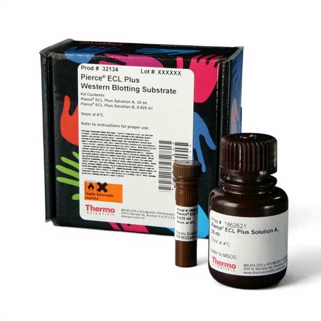
Pierce™ ECL Plus Western Blotting Substrate
Thermo Scientific Pierce ECL Plus Substrate is an acridan-based chemiluminescent and chemifluorescent HRP substrate for western blot detection using X-rayRead more
Have Questions?
Change view

| Catalog Number | DimensionsLxWxH |
|---|---|
| 32134 | |
| 32132 | |
| 32132X3 |
Catalog number 32134
Price (USD)
132.65
Online Exclusive
149.00Save 16.35 (11%)
Each
Price (USD)
132.65
Online Exclusive
149.00Save 16.35 (11%)
Each
Thermo Scientific Pierce ECL Plus Substrate is an acridan-based chemiluminescent and chemifluorescent HRP substrate for western blot detection using X-ray film or CCD- or laser-based imagers. This product is sold as Pierce ECL 2 Substrate (Cat. #PI80196X3) through Fisher Scientific and other channels.
Features of ECL Plus Substrate:
• Easy to use—can be substituted for the discontinued GE ECL Plus Substrate without any re-optimization
• Higher sensitivity—detect targets down to the low-picogram level
• Longer signal duration—sustained light output for as long as 5 hours
• More imaging options—X-ray, CCD or laser-based imagers
• More affordable—high quality and performance at a lower price than other suppliers' similar substrates
Pierce ECL Plus Substrate generates acridinium esters when it reacts with HRP. As these ester intermediates react with peroxide, they produce a strong and sustained chemiluminescent signal that can be captured by X-ray film CCD imaging. The substrate also produces a robust fluorescent signal that can be captured with fluorescence imagers. Pierce ECL Plus Substrate enables the detection of low-picogram amounts of target protein on nitrocellulose or PVDF membrane when probed with appropriate primary and secondary antibody concentrations. Its broad dynamic range, high sensitivity and long-lasting signal make Pierce ECL Plus Substrate an excellent choice for western blot analysis.
WARNING: Cancer – www.P65Warnings.ca.gov
For Research Use Only. Not for use in diagnostic procedures.
Specifications
Kit ContentsDetection Reagent A, 25 mL, Detection Reagent B, 0.625 mL
Recommended Antibody Concentrations2°: 1:25-200K (5-40ng/mL), 1°: 1:1K (0.05- 1μg/mL)
SensitivityLow picogram to mid-femtogram
Signal Duration5 hr.
Sufficient For250 cm2 of membrane
Detection MethodChemiluminescence, Fluorescence
For Use With (Application)Western Blot
FormLiquid
Membrane CompatibilityNitrocellulose, PVDF
Product LinePierce
Product TypeSubstrate
Quantity25 mL Kit
SubstrateHorseradish Peroxidase
Unit SizeEach
Contents & Storage
Upon receipt store at 4°C. Product shipped on ice.