Search


Citations & References (5)
Invitrogen™
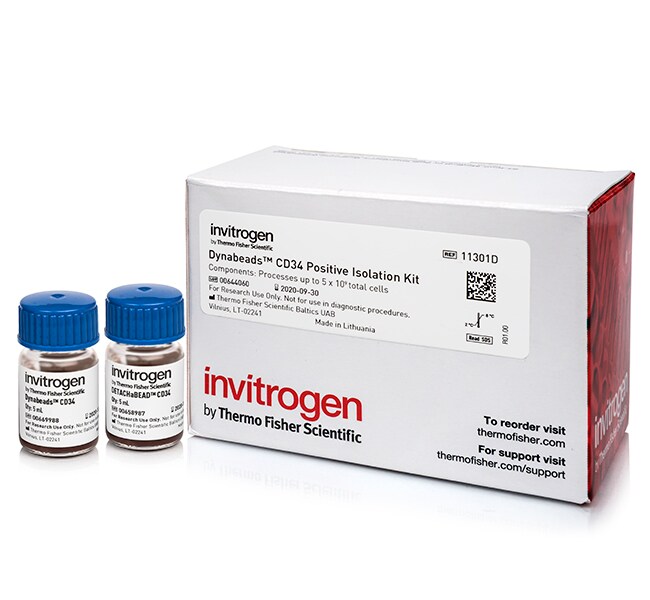
Dynabeads™ CD34 Positive Isolation Kit
Use this kit to isolate a high purity and yield of human CD34+ progenitor stem cells. Positively isolated cells areRead more
| Catalog Number | Quantity |
|---|---|
| 11301D | 5 mL |
Catalog number 11301D
Price (CNY)
12,587.00
飞享价
Ends: 31-Dec-2026
17,178.00Save 4,591.00 (27%)
Each
Quantity:
5 mL
Price (CNY)
12,587.00
飞享价
Ends: 31-Dec-2026
17,178.00Save 4,591.00 (27%)
Each
Use this kit to isolate a high purity and yield of human CD34+ progenitor stem cells. Positively isolated cells are bead and antibody-free, phenotypically unaltered and suitable for any downstream applications including flow cytometry, functional studies, and cell culture to produce dendritic cells (DC's). The Dynal™ CD34 Progenitor Cell Isolation System can also be used to isolate CD34+ cells from rhesus monkey (Macaca mulatta).
• Positive isolation of human CD34+ progenitor stem cells with bead release
• Stem cells can be isolated directly from whole blood, cord blood or bone marrow
• Isolated CD34+ cells can be used in any application, i.e. be derived into dendritic cells (DC's)
Starting samples:
PBMC from mobilized peripheral blood, bone marrow or cord blood.
For Research Use Only. Not for use in diagnostic procedures.
Specifications
Cell TypeStem Cells, Stem Cells (Hematopoietic)
Isolation TechnologyPositive Isolation
No. of CellsProcesses ∼5 x 10⁹ cells total
Output Viability>95%
Product LineDETACHaBEAD, DYNAL, Dynabeads
Purity or Quality GradeResearch Grade
Quantity5 mL
Sample TypeBlood (Mobilized Peripheral), Blood (Umbilical Cord), Bone Marrow
Shipping ConditionRoom Temperature
Starting Material Cell No.1 x 108 PBMCs per isolation
Target SpeciesHuman
Product TypePositive Cell Isolation Kit
Unit SizeEach
Contents & Storage
Each kit contains 5 mL Dynabeads™ coated with anti-CD34 monoclonal antibody, and 5 mL DETACHaBEAD™ CD34.
The kit can process up to 5 x 10⁹ cells. Store components at 2°C to 8°C.
The kit can process up to 5 x 10⁹ cells. Store components at 2°C to 8°C.
Frequently asked questions (FAQs)
My Dynabeads magnetic beads are not pelleting well with the magnet. Do you have any suggestions for me?
I have a long double-stranded DNA fragment I would like to isolate. What product do you recommend?
Can I use Dynabeads magnetic beads to isolate single-stranded DNA templates?
What is the magnetic susceptibility for Dynabeads magnetic beads?
How can I determine coupling efficiency of Dynabeads magnetic beads?
Citations & References (5)
Citations & References
Abstract
Gene expression analysis of hematopoietic progenitor cells identifies Dlg7 as a potential stem cell gene.
Journal:Stem Cells
PubMed ID:17322106
'Inducible hematopoietic stem/progenitor cell lines represent a model for studying genes involved in self-renewal and differentiation. Here, gene expression was studied in the inducible human CD34+ acute myelogenous leukemia cell line KG1 using oligonucleotide arrays and suppression subtractive cloning. Using this approach, we identified Dlg7, the homolog of the Drosophila
Infection of human CD34+ progenitor cells with Bartonella henselae results in intraerythrocytic presence of B. henselae.
Journal:Blood
PubMed ID:15860668
'Although there is evidence that endothelial cells are important targets for human pathogenic Bartonella species, the primary niche of infection is unknown. Here we elucidated whether human CD34+ hematopoietic progenitor cells (HPCs) internalize B. henselae and may serve as a potential niche of the pathogen. We showed that B. henselae
I branching formation in erythroid differentiation is regulated by transcription factor C/EBP{alpha}.
Journal:Blood
PubMed ID:17855628
'The histo-blood group i and I antigens have been characterized as straight and branched repeats of N-acetyllactosamine, respectively, and the conversion of the straight-chain i to the branched-chain I structure on red cells is regulated to occur after birth. It has been demonstrated that the human I locus expresses 3
Early lymphoid progenitors in mouse and man are highly sensitive to glucocorticoids.
Journal:Int Immunol
PubMed ID:15746243
Glucocorticoids are extensively used in anti-inflammatory therapy and may contribute to the normal regulation of lymphopoiesis. This study utilized new information about the early stages of lymphopoiesis in mouse and man to determine precisely which cell types are hormone sensitive. Cycling B lineage precursors were depleted in dexamethasone-treated mice, while
T cells modulate Epstein-Barr virus latency phenotypes during infection of humanized mice.
Journal:
PubMed ID:24390326
Human B cells, the main target of Epstein-Barr virus (EBV), can display several types of latent viral protein expression, denoted 0, I, IIa, IIb, or III. Of these, only type III expression induces proliferation of cells in vitro. These latency types are present at specific stages of infection and are