Search

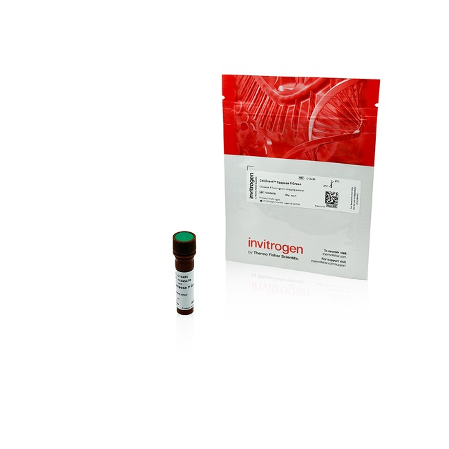
Invitrogen™
CellEvent Caspase 8 and 9 Detection Reagents
CellEvent™ Caspase Detection Reagents are fluorogenic, cell-permeant substrates for detecting activated caspase-3/7, caspase-8, and caspase-9 enzymes in live cells. No-wash formats support endpoint, kinetic, high-content imaging, and microplate apoptosis assays.
Have Questions?
Change view

| Catalog Number | Enzyme | Quantity |
|---|---|---|
| C10485 | Caspase 9 | 1 x 300 μL Vial |
| C10483 | Caspase 8 | 1 x 300 μL Vial |
| C10484 | Caspase 8 | 5 x 300 μL Vials |
| C10486 | Caspase 9 | 5 x 300 μL Vials |
| C10487 | Caspase 8 and Caspase 9 | 2 x 300 μL Vials |
Catalog number C10485
Price (CNY)
2,950.00
Each
Enzyme:
Caspase 9
Quantity:
1 x 300 μL Vial
Price (CNY)
2,950.00
Each
CellEvent™ Caspase Detection Reagents are cell-permeant, fluorogenic substrates for detecting activated caspase-3/7, caspase-8, and caspase-9 during apoptosis. Following cleavage by the target caspase, the released dye binds DNA and produces bright nuclear fluorescence in apoptotic cells. The no-wash reagents support live-cell and fixed-cell fluorescence imaging, high-content analysis, and microplate reader-compatible assays. They can be used for endpoint, kinetic, continuous-culture, and multiplexed workflows in 2D or 3D cell models.
CellEvent Caspase-8 and CellEvent Caspase-9 Green Detection Reagents are cell-permeant, fluorogenic substrates for detecting activated initiator Caspases during apoptosis. Caspase-8 is generally activated downstream of death receptor signaling and Caspase-9 is generally activated through the mitochondrial apoptosome pathway. Measuring these enzymes can provide information about upstream extrinsic and intrinsic apoptosis signaling, while recognizing that pathway crosstalk and cell-specific responses can influence Caspase activity.
Product features
- Detect activated Caspase-8 or Caspase-9 in living cells
- Produce bright green nuclear fluorescence for image-based identification and segmentation of Caspase-positive cells
- Use an add-and-read workflow with no cell lysis, permeabilization, or wash step required
- Support endpoint, kinetic, continuous-culture, high-content imaging, and microplate workflows
- Retain activated signal after formaldehyde-based fixation
- Support multiplex analysis with spectrally compatible cell-health and viability probes
Substrate specifics and principles of action
Each reagent contains a four-amino acid peptide recognition sequence linked to a fluorogenic DNA-binding dye. CellEvent Caspase-8 Green Detection Reagent uses the IETD sequence, while CellEvent Caspase-9 Green Detection Reagent uses the LEHD sequence. In cells without activated target Caspase, the intact reagent remains nonfluorescent because the peptide prevents the dye from binding DNA.
Unlike other caspase detection reagents that bind a single dye molecule to an activated enzyme in an irreversible reaction, the fluorogenic induction of CellEvent dye by cleavage does not inhibit caspase enzymes in the cell enabling signal amplification as the dye binds DNA producing bright green nuclear fluorescence response in cells with Caspase activity.
The fluorescence can be detected by microscopy, automated high-content imaging, or a compatible microplate reader. Imaging helps provide single-cell context, while microplate readers provide a population-level measurement. Whole-well assays may require optimization of cell density, treatment conditions, and the proportion of Caspase-positive cells to generate sufficient signal.
CellEvent Caspase reagent formats and portfolio
The CellEvent portfolio also includes reagents for measuring downstream executioner Caspase activity. CellEvent Caspase-3/7 Red Detection Reagents (C10430, C10431) provide a red nuclear signal for experiments multiplexing with GFP or compatible green-fluorescent markers. CellEvent Caspase-3/7 Green ReadyProbes™ Reagent (R37111) is supplied in a ready-to-use dropper bottle for a simple labeling workflow. Other CellEvent Caspase-3/7 Green formats include C10723, C10423, C10432, and C10433.
Assay versatility for live-cell imaging and endpoint analysis
CellEvent Caspase-8 and Caspase-9 reagents can be added directly to living cells in complete culture medium or a compatible physiological buffer. For endpoint analysis, cells are typically incubated with reagent for 3060 minutes at 37°C before measurement. The green reagents have excitation and emission maxima of approximately 502/530 nm and can be detected using standard FITC or GFP optical configurations.
For kinetic and continuous-culture studies, the reagent can remain in complete medium during extended culture of 2D or 3D models. Time-lapse imaging with an environmentally controlled microscope or on-stage incubator can monitor the appearance, timing, and distribution of Caspase-positive nuclei during prolonged treatment. The workflow is compatible with systems such as the EVOS™ M7000 Imaging System equipped with the EVOS Onstage Incubator, or similarly configured microscopes with appropriate environmental control.
For microplate-based high-content imaging, the bright nuclear signal supports automated segmentation and measurements such as nuclear intensity or percentage of positive cells. The fluorescence response can also be measured with a microplate reader for population-level analysis.
The reagents must be activated by Caspases in living cells and cannot be applied to previously fixed samples to generate signal. After sufficient cleavage and signal development, cells may be preserved with a formaldehyde-based fixative for endpoint imaging, permeabilization, or subsequent analysis with compatible fixed-cell reagents.
Format advantages
- Ready-to-dilute 100X aqueous concentrates at 500 μM
- Direct dilution into compatible physiological buffer or complete culture medium
- Storage at 28°C without preparation of a DMSO stock solution
- Detection with standard FITC or GFP optical configurations
Applications
- Drug discovery, compound profiling, and mechanism-of-action studies
- Live-cell and time-lapse fluorescence imaging
- On-stage incubator and continuous-culture experiments
- High-content screening and automated image analysis
- Microplate reader-based Caspase quantitation
- Fixed-cell endpoint imaging after live-cell activation
- Longitudinal studies in 2D and 3D cell models
Multiplex analysis of Caspase activity, mitochondrial function, and membrane integrity
For Research Use Only. Not for use in diagnostic procedures.
Specifications
AssayApoptosis
ColorGreen
Compatible CellsLive mammalian cells
EnzymeCaspase 9
Excitation/Emission502/530
For Use With (Application)Live cell imaging, 2D and 3D cell culture, continuous culture, drug discovery, apoptosis
For Use With (Equipment)Fluorescence Microscopes, High Content Instruments, Live Cell Imaging, Microplate Readers, Flow Cytometers
Formulation500 μM 100x aqueous concentrate solution
Incubation Time30 to 60 min. or Continuous Culture
No. of Reactions100 Tests
Product LineCellEvent
Product TypeCaspase detection reagent
Quantity1 x 300 μL Vial
ConjugateCellEvent Caspase 9 Green
Detection MethodFluorescence
FormatVial
Unit SizeEach
Contents & Storage
•1 vial 300 μL 100x aqueous concentrate Caspase 9 detection reagent
•Store at 2°C to 8°C
•Protect from light
•Store at 2°C to 8°C
•Protect from light