Search


Citations & References (7)
Invitrogen™
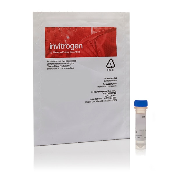
Fluo-5N, Pentapotassium Salt, cell impermeant
Analog of fluo-4 with lower Ca2+-binding affinity, making it suitable for detecting intracellular calcium levels in 1μM to 1 mM range that would saturate response of fluo-3 and fluo-4
| Catalog Number | Quantity |
|---|---|
| F14203 also known as F-14203 | 500 μg |
Catalog number F14203
also known as F-14203
Price (CNY)
5,142.00
Each
Quantity:
500 μg
Price (CNY)
5,142.00
Each
Labeled calcium indicators are molecules that exhibit an increase in fluorescence upon binding Ca2+. Fluo-5F, fluo-5N, and fluo-4ff are analogs of fluo-4 with lower Ca2+-binding affinity, making them suitable for detecting intracellular calcium levels in the 1 μM to 1 mM range that would saturate the response of fluo-3 and fluo-4. Cells may be physically loaded with the cell-impermeant salt forms of these indicators using patch pipette or microinjection or our Influx™ pinocytotic cell-loading reagent (Cat. No. I14402). These indicators are compatible with excitation at 488 nm by argon-ion laser sources, making them useful for confocal microscopy, flow cytometry, and microplate screening applications.
Calcium Indicator (Cell-Impermeant Salts) Specifications:
- Label (Ex/Em of Ca2+–bound form): Fluo-5N (494/516 nm)
- Fluorescence intensity increase upon binding Ca2+: >100 fold
- Kd for Ca2+ in buffer: ∼90 μM
- Exhibit fluorescence increase upon binding Ca2+ with little shift in wavelength
Using TPEN to Control Heavy Metal Cations
In addition, BAPTA-based indicators such as these bind various heavy metal cations (e.g., Mn2+, Zn2+, Pb2+) with substantially higher affinity than Ca2+. Perturbations to calcium measurements caused by presence of these ions can be controlled using the heavy metal-selective chelator (Cat. No. TPEN).
More Choices for Fluorescent Calcium Indicators
We offer a large selection of Molecular Probes™ calcium indicators for use in various experimental scenarios. For more information, review Fluorescent Ca2+ Indicators Excited with Visible Light—Section 19.3 in the Molecular Probes™ Handbook.
For UV-excitable Ca2+ indicators, protein-based Ca2+ indicators, conjugates of Ca2+ indicators, and for fluorescence-based indicators of other metal ions (i.e., Mg2+, Zn2+) review Indicators for Ca2+, Mg2+, Zn2+ and Other Metal Ions—Chapter 19 in the Molecular Probes™ Handbook.
For Research Use Only. Not for human or animal therapeutic or diagnostic use.
For Research Use Only. Not for use in diagnostic procedures.
Specifications
Detection MethodFluorescence
Dye TypeFluo-5N
Quantity500 μg
Shipping ConditionRoom Temperature
Emission Wavelength Range516 nm
Excitation Wavelength Range494 nm
For Use With (Application)Calcium Indicator
For Use With (Equipment)Confocal Microscope, Fluorescence Microscope, High Content Analysis Instrument, HTS Reader, Microplate Reader, Fluorescent Imager
Product TypeCalcium Indicator
Unit SizeEach
Contents & Storage
Store in freezer -5°C to -30°C and protect from light.
Citations & References (7)
Citations & References
Abstract
Rational design of protein-based MRI contrast agents.
Journal:J Am Chem Soc
PubMed ID:18576649
We describe the rational design of a novel class of magnetic resonance imaging (MRI) contrast agents with engineered proteins (CAi.CD2, i = 1, 2,..., 9) chelated with gadolinium. The design of protein-based contrast agents involves creating high-coordination Gd(3+) binding sites in a stable host protein using amino acid residues and
Concurrent maturation of inner hair cell synaptic Ca2+ influx and auditory nerve spontaneous activity around hearing onset in mice.
Journal:
PubMed ID:23804089
Hearing over a wide range of sound intensities is thought to require complementary coding by functionally diverse spiral ganglion neurons (SGNs), each changing activity only over a subrange. The foundations of SGN diversity are not well understood but likely include differences among their inputs: the presynaptic active zones (AZs) of
Low-affinity Ca2+ indicators compared in measurements of skeletal muscle Ca2+ transients.
Journal:Biophys J
PubMed ID:19804716
The low-affinity fluorescent Ca(2+) indicators OGB-5N, Fluo-5N, fura-5N, Rhod-5N, and Mag-fluo-4 were evaluated for their ability to accurately track the kinetics of the spatially averaged free Ca(2+) transient (Delta[Ca(2+)]) in skeletal muscle. Frog single fibers were injected with one of the above indicators and, usually, furaptra (previously shown to rapidly
Tuning of synapse number, structure and function in the cochlea.
Journal:Nat Neurosci
PubMed ID:19270686
Cochlear inner hair cells (IHCs) transmit acoustic information to spiral ganglion neurons through ribbon synapses. Here we have used morphological and physiological techniques to ask whether synaptic mechanisms differ along the tonotopic axis and within IHCs in the mouse cochlea. We show that the number of ribbon synapses per IHC
Confocal imaging of CICR events from isolated and immobilized SR vesicles.
Journal:Cell Calcium
PubMed ID:16122794
The Ca2+ concentration inside the sarcoplasmic reticulum ([Ca2+]SR) is a difficult parameter to measure in ventricular cardiac myocytes. Interference from Ca2+-sensitive dye loading into cellular compartments other than the SR interferes with free Ca2+ measurement. In addition, the composition of the cytosol surrounding the SR in intact cells cannot be