Search

迎接更强大的成像能力
全自动 EVOS™ M7000 成像系统 专为简化最具挑战性的载玻片和细胞成像应用而设计,涵盖 活细胞分析、图像拼接(tiling) 以及 Z 轴堆叠(Z-stacking) 等功能。
该系统配备了先进的 二维反卷积(2D deconvolution) 技术,可显著提升 信噪比(SNR),获得更加清晰、准确的图像信息,从而帮助研究人员发掘样品中原本难以观察到的关键信息和细节。
产品特点
除了所有 EVOS microscopes的通用功能外,EVOS M7000成像系统还提供:
全自动化
自动化流程有助于简化工作流程并提高实验的可重复性
速度
可在不到5分钟内扫描96孔板的3个荧光通道
二维去卷积
捕捉具有卓越清晰度和细节的图像
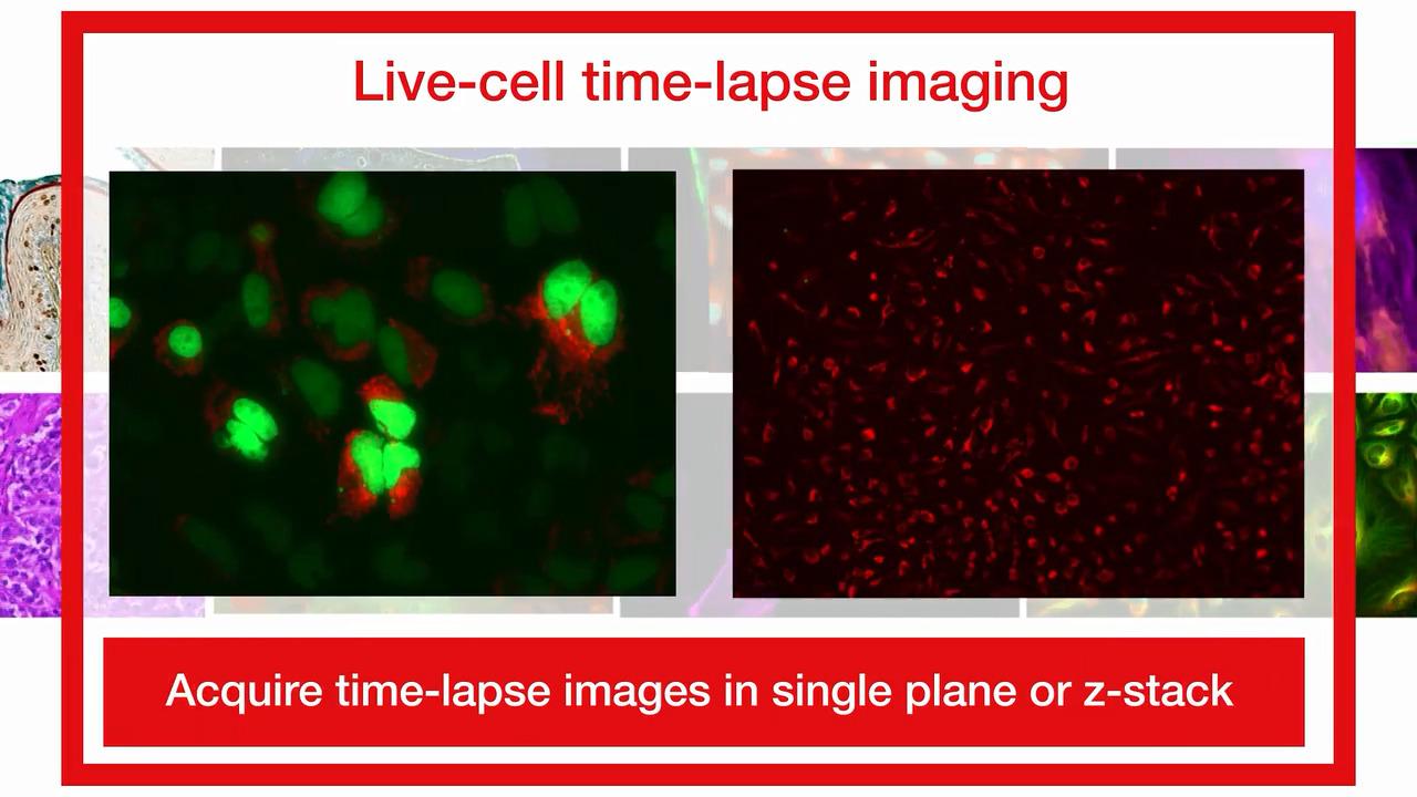
延时活细胞成像
可选配的台式培养箱能够精确控制温度、湿度和气体浓度
区域视图
可在单视野模式与低倍、高倍扫描模式之间快速无缝切换,轻松定义并捕获感兴趣区域
数据分析
可将图像无缝传输至可选的 Celleste 图像分析软件,获取强大的图像分割、分类及基于细胞的分析工具
Virtual 360º view
虚拟体验 EVOS M7000 成像系统
Explore 360º view

高级二维去卷积
体验二维去卷积带来的卓越清晰度和细节

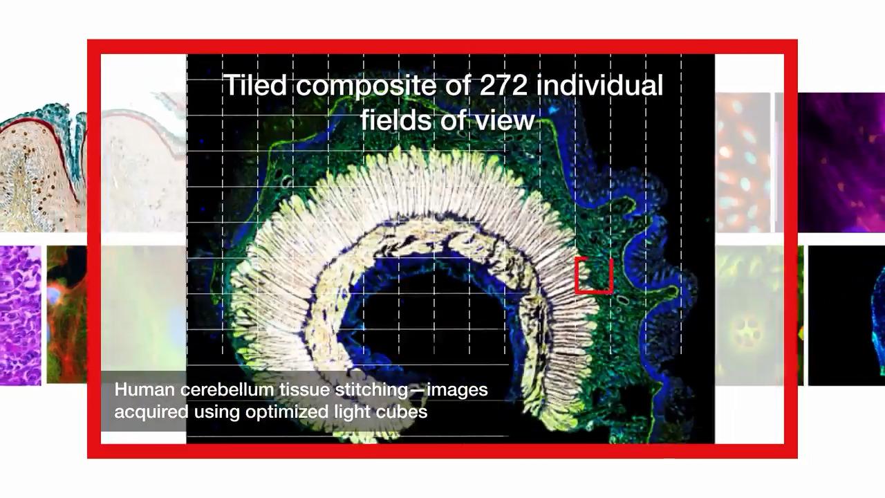
FluoCells™ 预制切片 #3(小鼠肾组织切片,采用 Alexa Fluor™ 488 WGA、Alexa Fluor™ 568 Phalloidin 和 DAPI 染色),使用 EVOS™ M7000 成像系统 在 4× 物镜 下成像,并通过 EVOS Analysis 软件进行反卷积处理(货号 AMEP4922) 。

U2OS 细胞 接种于 Nunc™ 玻底培养皿(货号 150682)中,并分别采用以下试剂进行标记:Hoechst 33342(三盐酸三水合物,10 mg/mL) (Cat. No. H3570, 细胞核—蓝色), Alexa Fluor™ 488 phalloidin (货号 A12379, 肌动蛋白—绿色), α-微管蛋白单克隆抗体 (236-10501) (货号 A11126) 并使用 Alexa Fluor™ 647 交叉吸附型山羊抗小鼠 IgG(H+L)二抗(货号 A21235, 微管—紫色). 使用 EVOS M7000 成像系统配备 Olympus 100× 半复消色差物镜(0.95 NA / 0.2 WD) (货号 AMEP4988)进行成像。

Hela 细胞, 经 PFA 固定并用 DAPI、抗 coilin(GFP)、抗微管蛋白(Cy5)和 Ki-67(RFP)染色, 使用 EVOS M7000 成像系统 with 20x fluorite objective (Cat. No. AMEP4698), 并通过 EVOS Analysis 软件进行去卷积处理
EVOS M7000 HCA 套装
EVOS M7000 成像系统高内涵分析套装 (AMF7000HCA) 将全自动倒置式多通道荧光和透射光成像系统 with Celleste 6 Image Analysis Software, a full-featured image analysis suite.Celleste 6 图像分析软件便于用户从低通量过渡到高内涵分析(HCA),同时保持 EVOS M7000 自动显微镜的多功能性和强大性能。
通过基于板的多通道分析(MCA)协议,结合基于机器学习的算法和图标驱动的向导式工作流程,Celleste 6 软件可帮助您高效地对各种检测或生物应用的图像进行分割和分类,包括神经突生长、血管生成、细胞活力、转染效率等。该软件还提供多种孔板数据展示方式,包括热图、图像拼贴和动力学曲线选项。
EVOS M7000 & Celleste 6 HCA 套装列表
描述 |
货号 |
|---|---|
EVOS™ M7000 成像系统,高内涵分析(HCA)套装
|
|
EVOS™ M7000 成像系统,高内涵分析(HCA)套装(含 3D 功能)
|

精选图像与视频


技术规格
| 类别 | 属性 | 描述 |
| 光学系统 | 光学描述 | 无限远校正光学系统;RMS 螺纹物镜,45 mm 齐焦距离 |
| 成像模式 | 荧光、明场、彩色明场、相差 | |
| 成像方法 | 单色、多色、区域扫描(拼接/平铺)、时间推移(time-lapse)、Z 轴堆叠、视频采集 | |
| 照明 | 可调强度 LED 光源模块,集成硬涂层滤光片组,使用寿命 >50,000 小时 | |
| 光源模块容量 | 5 位仓位:4 个荧光光源模块 + 1 个明场 | |
| 光源模块(不含) | 提供多种标准及特殊光源模块,常用激发/发射(ex/em): • DAPI (357/447 nm) • GFP (470/525 nm) • RFP (531/593 nm) • Texas Red (585/624 nm) • Cy5 (628/692 nm) |
|
| 物镜容量 | 5 位自动旋转物镜转盘 | |
| 物镜(不含) | 多种高质量长工作距离(LWD)及盖玻片校正物镜可选 | |
| 聚光镜 | 60 mm 长工作距离聚光镜;4 位转盘,含透明孔径和 3 个相差环 | |
| 对焦机构 | 自动对焦,亚微米级分辨率(0.150 µm 单步精度) | |
| 单色相机 | 高灵敏度 3.2 MP(2048 × 1536)CMOS 传感器,像素尺寸 3.45 µm | |
| 彩色相机 | 高灵敏度 3.2 MP(2048 × 1536)CMOS 传感器,像素尺寸 3.45 µm | |
| 捕获格式 | 16 位 RAW 单色: TIFF、PNG(12 位动态范围) 8 位彩色:TIFF、PNG、JPG 视频/延时成像:AVI、WMV |
|
| 载物台 | 电源 | 24V 交流适配器,配备各国标准电源线 |
| 尺寸(宽 × 高 × 深) | 18 x 14 x 13 英寸 45.7 x 33.0 x 35.6 cm |
|
| 重量 | 26 镑 (11.8 kg) | |
| X/Y 扫描台 | 行程 120 mm × 80 mm,亚微米分辨率 可直接放入的样品插块,用于放置培养皿/多孔板,并在长时间扫描中固定样品 |
|
| 台体机构 | 电动 | |
| 自动化 | 板扫描 | 多孔板自动扫描 |
系统 网络 |
计算机 | 外置 Dell XE4 计算机:Intel® Core™ i9 处理器,128 GB DDR4 内存,2 TB SSD,NVIDIA® Quadro® A4000(8 GB 独立显卡),运行 Windows® 10 21H2 LTSC |
| 输出接口 | 计算机接口:1 × USB 3.1 Gen 2 Type-C;5 × USB 3.1 Gen 1 Type-A;4 × USB 2.0 Type-A;1 × 串口;2 × DisplayPort 1.2;1 × RJ-45;2 × PS/2;1 × UAJ;1 × Line-out | |
| 网络能力 | 通过以太网电缆连接 Windows/SMB 网络 |
Ordering information
For Research Use Only. Not for use in diagnostic procedures.