Search

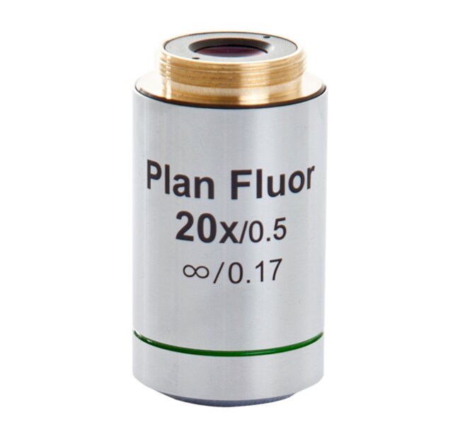
EVOS™ 20X 物镜,萤石,盖玻片校正
This fluorite objective is ideal for fluorescence and demanding transmitted-light applications. This is a coverslip-corrected objective that is optimized for了解更多信息
| 货号 | 数量 |
|---|---|
| AMEP4698 | 各 1 个 |
货号 AMEP4698
价格(CNY)
-
数量:
各 1 个
This fluorite objective is ideal for fluorescence and demanding transmitted-light applications. This is a coverslip-corrected objective that is optimized for imaging through #1.5 coverslips. All EVOS objectives offer outstanding optical performance from visible light to near infrared light. The extensive choice of objectives satisfies needs across the spectrum of magnifications and optical specifications.
Additional characteristics of this EVOS objective:
• Magnification: 20X
• Numerical Aperture: 0.50
• Working Distance: 2.5 mm
Image Quality
Microscope objectives may be the most important components of an optical microscope because they are responsible for primary image formation. Image quality is crucial to experimental success and a requirement for publication; EVOS objectives afford that quality across the visible spectrum to near infrared light. This performance results from years of lens manufacture perfection. EVOS objectives have the same or better numerical apertures as any other manufacturer's in the same class, and the broad selection means you have choices for your imaging requirements.
Objective Classes
Achromat objectives are perfect for general applications, with standard correction of color and focus.
Fluorite objectives deliver excellent resolution and are made with higher numerical apertures than achromat objectives, resulting in brighter fluorescence signal and higher contrast imaging. The higher optical quality greatly reduces optical aberrations, and corrections for color and focus are at higher levels than achromat objectives. Fluorite objectives are ideally suited for fluorescence and demanding transmitted light applications, where the higher contrast make them ideally suited for color imaging.
Apochromat objectives are manufactured to the highest levels of resolution, fluorescence brightness, and contrast; chromatic aberrations are almost eliminated. They are recommended for the most demanding applications, particularly at magnifications of 60x and above. Apochromatic objectives are the best choice for the capture of color images in white light.
Brightfield Contrast versus Phase Contrast Objectives
Brightfield is the most basic form of light microscopy and is accomplished by sample absorption of light. A higher density area in a sample will absorb more light, thus increasing contrast in those areas.
Phase contrast objectives are most useful for hard to see, translucent specimens. This method of contrast is accomplished by converting phase shifts, caused by light passing through a translucent specimen, into brightness changes (i.e., contrast).
Long Working Distance versus Coverslip-Corrected Objectives
Long working distance (LWD) objectives are optimized for use through vessels with a nominal wall thickness of 0.9-1.5 mm. This includes vessels commonly used in cell culture and cell-based assays, such as slides, cell culture dishes and flasks, microtiter well plates, etc. Coverslip-corrected objectives are optimized for use through #1.5 coverslips (thickness approximately 0.17 mm). These objectives have a higher magnification/NA ratio and provide higher resolution compared to LWD objectives.
For additional choices, visit the EVOS Objectives selection guide
Explore the entire EVOS line of imaging systems and accessories
仅供科研使用。不可用于诊断程序。
规格
焦距2.5 mm
适用于(设备)EVOS™ XL Core 成像系统、EVOS™ FL 彩色成像系统、EVOS™ XL 成像系统、EVOS™ FL 成像系统、EVOS™ FL 自动成像系统
透镜类型平场萤石
放大20X
数值孔径0.50
数量各 1 个
产品线EVOS:
类型物镜
Unit SizeEach