Search


Citations & References (8)
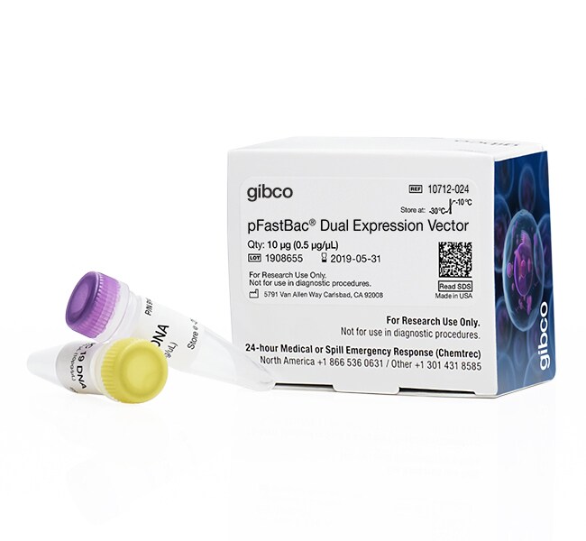
Gibco™
pFastBac™ Dual Expression Vector
The pFastBac™ Dual vector features two promoters in a single vector for expression of two proteins simultaneously in insect cellsRead more
| Catalog Number | Quantity |
|---|---|
| 10712024 | 10 μg |
Catalog number 10712024
Price (CNY)
12,962.00
Each
Quantity:
10 μg
Price (CNY)
12,962.00
Each
The pFastBac™ Dual vector features two promoters in a single vector for expression of two proteins simultaneously in insect cells when using the Bac-to-Bac® Baculovirus Expression System (Cat. No. 10359-016). The vector has two strong promoters, the polyhedrin promoter and the p10 promoter, for high-level expression. An expression control is included that expresses both CAT and β-glucuronidase (gus).
For Research Use Only. Not for use in diagnostic procedures.
Specifications
Product TypeExpression Vector
Quantity10 μg
VectorpFastBac
Cloning MethodRestriction Enzyme/MCS
Product LinepFastBac
Promoterp10, Polyhedrin
Protein TagUntagged
Unit SizeEach
Contents & Storage
pFastBac™ Dual (10 μg) and pFastBac™ Dual Control vector (4 ng) are supplied lyophilized.
Store at -20°C. Vectors are guaranteed stable for 6 months when properly stored.
Store at -20°C. Vectors are guaranteed stable for 6 months when properly stored.
Frequently asked questions (FAQs)
I cannot grow this white colony in liquid culture. What should I do?
What has happened when I see blue colonies? How about colonies which are blue in the center and white on the edges?
I'm getting mostly white/wild-type plaques instead of blue/recombinant plaques. What am I doing wrong?
I've infected my cells and see large polyhedra in one cell and smaller polyhedra (more numerous) in a neighboring cell. Is this normal?
I'm worried that I am not getting plaques. How many days does it take to see plaques and what size are they typically?
Citations & References (8)
Citations & References
Abstract
Molecular characterization of hCTR1, the human copper uptake protein.
Journal:J Biol Chem
PubMed ID:12034741
'We have expressed hCTR1, the human copper transporter, in Sf9 cells using a baculovirus-mediated expression system, and we observed greatly enhanced copper uptake. Western blots showed that the protein is delivered to the plasma membrane, where it mediates saturable copper uptake with a K(m) of approximately 3.5 microm. We also
Structure of the reovirus membrane-penetration protein, Mu1, in a complex with is protector protein, Sigma3.
Journal:Cell
PubMed ID:11832217
Cell entry by nonenveloped animal viruses requires membrane penetration without membrane fusion. The reovirus penetration agent is the outer-capsid protein, Mu1. The structure of Mu1, complexed with its
Molecular characterization of major cat allergen Fel d 1: expression of heterodimer by use of a baculovirus expression system.
Journal:J Biol Chem
PubMed ID:15546862
Fel d 1 is a major cat allergen inducing allergic rhinitis and asthma in sensitized individuals. It has a more complex structure when compared with other allergens and therefore expression of recombinant Fel d 1 has been considered a challenge. The present study shows for the first time that a
A novel 43-kDa protein as a negative regulatory component of phenoloxidase-induced melanin synthesis.
Journal:J Biol Chem
PubMed ID:15857824
The melanization reaction induced by activated phenoloxidase in arthropods is important in the multiple host defense innate immune reactions, leading to the sequestration and killing of invading microorganisms. This reaction ought to be tightly controlled because excessive formation of quinones and systemic hypermelanization are deleterious to the hosts, suggesting that
Human MutY homolog, a DNA glycosylase involved in base excision repair, physically and functionally interacts with mismatch repair proteins human MutS homolog 2/human MutS homolog 6.
Journal:J Biol Chem
PubMed ID:11801590
Adenines mismatched with guanines or 7,8-dihydro-8-oxo-deoxyguanines that arise through DNA replication errors can be repaired by either base excision repair or mismatch repair. The human MutY homolog (hMYH), a DNA glycosylase, removes adenines from these mismatches. Human MutS homologs, hMSH2/hMSH6 (hMutSalpha), bind to the mismatches and initiate the repair on