Search


Citations & References (15)
Invitrogen™
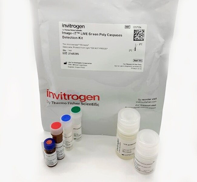
Image-iT™ LIVE Red and Green Caspase Apoptosis Detection Kits for microscopy
Detect apoptosis via microscopy, including fluorescence microscopy and HCS, with Image-iT LIVE Red and Green Caspase Apoptosis Detection Kits that report poly and caspase-3/7 activities.
Have Questions?
Change view

| Catalog Number | Color | Enzyme | Label or Dye |
|---|---|---|---|
| I35102 | Red, Green, Blue | Caspase 3/7 | SR-DEVD-FMK, SYTOX Green, Hoechst 33342 |
| I35101 | Red, Green, Blue | Poly Caspases | SR-VAD-FMK, SYTOX Green, Hoechst 33342 |
| I35104 | Green, Red, Blue | Poly Caspases | FAM-VAD-FMK, Propidium Iodide, Hoechst 33342 |
| I35106 | Green, Red, Blue | Caspase 3/7 | FAM-DEVD-FMK, Propidium Iodide, Hoechst 33342 |
Catalog number I35102
Price (CNY)
7,136.00
Each
Color:
Red, Green, Blue
Enzyme:
Caspase 3/7
Label or Dye:
SR-DEVD-FMK, SYTOX Green, Hoechst 33342
Price (CNY)
7,136.00
Each
Detect and evaluate caspase activation, membrane permeability, and cell cycle using Image-iT LIVE red and green poly and caspase-3 and-7 apoptosis detection kits for microscopy, including fluorescence microscopy and high content screening (HCS). The Image-iT LIVE red and green caspase detection kits use fluorescent inhibitor of caspases (FLICA) methodology to detect and report caspase activity, a measure of cell apoptosis. Specifically, the Image-iT LIVE red and green caspase detection kits contain the substrates SR-VAD-FMK and SR-DEVD-FMK (for red fluorescence), or FAM-VAD-FMK and FAM-DEVD-FMK (green fluorescence), for poly caspases and caspase-3/7 detection, respectively. They also include Hoechst 33342 and propidium iodide stains, enabling simultaneous evaluation of membrane permeability and cell cycle by microscopy and HCS.
The Image-iT LIVE red and green caspase detection kits for microscopy employ a novel approach to detecting active caspases: both assay kits take advantage of a fluorescent inhibitor of caspases (FLICA) methodology, which is an affinity label. This reagent associates a fluoromethyl ketone (FMK) moiety, which can react covalently with a cysteine, with a caspase-specific amino acid sequence. Different amino acid recognition sequences are used to detect different caspases: valine-alanine-aspartic acid (VAD) for poly-caspases (including caspase-1, -3, -4, -5, -6, -7, -8, and -9), and aspartic acid-glutamic acid-valine-aspartic acid (DEVD) for caspase 3/7. A sulforhodamine (SR) group or carboxyfluorescein (FAM) group is attached as a reporter.
The FLICA reagent is thought to interact with the enzymatic reactive center of an activated caspase via the recognition sequence, and then to attach covalently through the FMK moiety. The FLICA inhibitor is cell permeant and noncytotoxic. Unbound FLICA molecules diffuse out of the cell and are washed away; the remaining red or green fluorescent signal is a direct measure of the amount of active caspase present at the time the inhibitor was added. The approximate excitation and emission peaks of the FLICA, propidium iodide, and Hoechst 33342 reagents are 488⁄ nm/530 nm, 535⁄ nm/6617 nm, and 350 nm/⁄461 nm, respectively. The detection reagents included within the Image-iT LIVE Red Poly Caspases, Red Caspase-3/7, Green Poly Caspases, and Green Caspase-3/7 Detection kits are SR-VAD-FMK, SR-DEVD-FMK, FAM-VAD-FMK, and FAM-DEVD-FMK, respectively.
The FLICA reagent is thought to interact with the enzymatic reactive center of an activated caspase via the recognition sequence, and then to attach covalently through the FMK moiety. The FLICA inhibitor is cell permeant and noncytotoxic. Unbound FLICA molecules diffuse out of the cell and are washed away; the remaining red or green fluorescent signal is a direct measure of the amount of active caspase present at the time the inhibitor was added. The approximate excitation and emission peaks of the FLICA, propidium iodide, and Hoechst 33342 reagents are 488⁄ nm/530 nm, 535⁄ nm/6617 nm, and 350 nm/⁄461 nm, respectively. The detection reagents included within the Image-iT LIVE Red Poly Caspases, Red Caspase-3/7, Green Poly Caspases, and Green Caspase-3/7 Detection kits are SR-VAD-FMK, SR-DEVD-FMK, FAM-VAD-FMK, and FAM-DEVD-FMK, respectively.
For Research Use Only. Not for use in diagnostic procedures.
Specifications
ColorRed, Green, Blue
DescriptionImage-iT LIVE Red Caspase-3 and -7 Detection Kit
EnzymeCaspase 3/7
Excitation/EmissionSR-DEVD-FMK: 550/595
SYTOX Green: 504/523
Hoechst 33342: 350/461
SYTOX Green: 504/523
Hoechst 33342: 350/461
For Use With (Equipment)Fluorescence Microscope
Label TypeOther Label(s) or Dye(s)
Label or DyeSR-DEVD-FMK, SYTOX Green, Hoechst 33342
No. of Reactions25 tests (labeling volumes of 300μL)
Product LineImage-iT
Product TypeCaspase 3/7 Reagent
Quantity1 kit
Shipping ConditionRoom Temperature
Detection MethodFluorescence
FormatSlide
Unit SizeEach
Contents & Storage
Store in refrigerator (2–8°C) and protect from light.
Citations & References (15)
Citations & References
Abstract
Endoplasmic Reticulum Stress and Apoptosis Contribute to the Pathogenesis of Dominantly Inherited Isolated GH Deficiency Due to GH1 Gene Splice Site Mutations.
Journal:
PubMed ID:23736291
Dominantly inherited isolated GH deficiency is mainly caused by a heterozygous donor site mutation of intron 3 in the GH1 gene. An exon 3 deletion in GH (del32-71 GH) is produced from a mutant allele, whereas wild-type GH is produced from the other allele. Several studies have demonstrated a dominant
Characterization of Differentiated SH-SY5Y as Neuronal Screening Model Reveals Increased Oxidative Vulnerability.
Journal:J Biomol Screen
PubMed ID:26738520
JUND regulates pancreatic ß cell survival during metabolic stress.
Journal:Mol Metab
PubMed ID:31023625
'In type 2 diabetes (T2D), oxidative stress contributes to the dysfunction and loss of pancreatic ß cells. A highly conserved feature of the cellular response to stress is the regulation of mRNA translation; however, the genes regulated at the level of translation are often overlooked due to the convenience of
CD95-mediated apoptosis in Burkitt's lymphoma B-cells is associated with Pim-1 down-regulation.
Journal:Biochim Biophys Acta Mol Basis Dis
PubMed ID:27641442
'B-cells of the high-grade non-Hodgkin lymphoma Burkitt''s lymphoma (BL) overexpress survival oncoproteins, including the proviral integration site for Moloney murine leukaemia virus kinase (Pim)-1, and become apoptosis resistant. Activated death receptor CD95 after ligation with anti-CD95 monoclonal antibody (mAb) resulted in the regression of BL via induction of apoptosis, suggesting
Impaired serotonin communication during juvenile development in rats diminishes adult sperm quality.
Journal:Syst Biol Reprod Med
PubMed ID:29788785
'Spermatogenesis and steroidogenesis are testicular functions regulated by gonadotrophins as well as other factors, including serotonin. Testicular serotonin acts as an autocrine regulator of testosterone secretion, but studies on its role in spermatogenesis and sperm quality are scarce. Here, we analyzed the effects of intratesticular inhibition of serotonin synthesis on