Search


Citations & References (29)
Invitrogen™
Calcium Phosphate Transfection Kit
The Calcium Phosphate Transfection Kit provides high-quality reagents to enable the introduction of DNA into eukaryotic cells via calcium phosphateRead more
| Catalog Number | Quantity |
|---|---|
| K278001 | 75 Reactions |
Catalog number K278001
Price (CNY)
8,788.00
Each
Quantity:
75 Reactions
Price (CNY)
8,788.00
Each
The Calcium Phosphate Transfection Kit provides high-quality reagents to enable the introduction of DNA into eukaryotic cells via calcium phosphate co-precipitation.
How it works
The calcium phosphate transfection method for introducing DNA into mammalian cells is based on forming a calcium phosphate-DNA precipitate. Calcium phosphate facilitates the binding of the DNA to the cell surface. DNA then enters the cell by endocytosis. The method was first developed by Graham and van der Ebb and was later modified by Wigler. The procedure is routinely used to transfect a wide variety of cell types for transient expression or for producing stable transformants. The DNA is mixed directly with a concentrated solution of CaCl2, which is then added dropwise to a phosphate buffer to form a fine precipitate. Aeration of the phosphate buffer while adding the DNA-CaCl2 solution helps to ensure that the precipitate that forms is as fine as possible, which is important because clumped DNA will not adhere to or enter the cell as efficiently.
For Research Use Only. Not for use in diagnostic procedures.
Specifications
For Use With (Application)Transfection
High-throughput CompatibilityNot High-throughput Compatible (Manual)
Product TypeTransfection Kit
Quantity75 Reactions
Serum CompatibleNo
Cell TypeEstablished Cell Lines
Format6-well Plate, 12-well Plate, 24-well Plate, 48-well Plate, 96-well Plate, Flasks
Sample TypePlasmid DNA
Transfection TechniqueChemical Transfection
Unit SizeEach
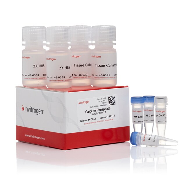
Contents & Storage
2 × 12 ml Tissue Culture Sterile Water
2 × 12 ml 2X HEPES Buffered Saline (HBS)
3 × 1 ml 2 M CaCl2
20 μg pcDNA™3.1/His/lacZ
Store all components at -5 to -30°C.
2 × 12 ml 2X HEPES Buffered Saline (HBS)
3 × 1 ml 2 M CaCl2
20 μg pcDNA™3.1/His/lacZ
Store all components at -5 to -30°C.
Frequently asked questions (FAQs)
I have tried several different transfection reagents and have failed to transfect my gene into my cell line of interest. Do you have any suggestions?
How do I perform a dose-response curve or kill curve?
What is the main advantage of viral transduction over transfection?
What is the main advantage of lipid-mediated transfection over calcium phosphate-mediated transfection?
What is the main difference between transient and stable transfection?
Citations & References (29)
Citations & References
Abstract
A Novel Homeobox Protein which Recognizes a TGT Core and Functionally Interferes with a Retinoid-responsive Motif
Journal:Nature
PubMed ID:1899916
'The synapsins are a family of closely related phosphoproteins (termed synapsins Ia, Ib, IIa and IIb) associated with synaptic vesicles and implicated in the short-term regulation of neurotransmitter release from nerve endings. During development, expression of the synapsins correlates temporally with synapse formation, but there has been no direct evidence
Suppression of cell transformation by the cyclin-dependent kinase inhibitor p57KIP2 requires binding to proliferating cell nuclear antigen.
Journal:Proc Natl Acad Sci U S A
PubMed ID:9465025
'Proper control of the mammalian cell cycle requires the function of cyclin-dependent kinase (CDK) inhibitors. The p21 family currently includes three distinct genes, p21, p27(Kip1), and p57(Kip2), that share a common N-terminal domain for binding to and inhibiting the kinase activity of CDK-cyclin complexes. The p21 protein also binds to
Ligand binding to macrophage scavenger receptor-A induces urokinase- type plasminogen activator expression by a protein kinase-dependent signaling pathway.
Journal:J Biol Chem
PubMed ID:9422792
'Macrophage scavenger receptor-type A (MSR-A) has been implicated in the transmission of cell signals and the regulation of diverse cellular functions (Falcone, D. J., and Ferenc, M. J. (1988) J. Cell. Physiol. 135, 387-396; Falcone, D. J., McCaffrey, T. A., and Vergilio, J. A. (1991) J. Biol. Chem. 266, 22726-22732;
A new cationic liposome reagent mediating nearly quantitative transfection of animal cells.
Journal:Biotechniques
PubMed ID:1867862
'One of the most efficient systems for the expression of genes in the cytoplasm of animal cells utilizes a recombinant vaccinia virus encoding the bacteriophage T7 RNA polymerase. Cells infected with this virus are transfected with plasmid DNAs containing the gene to be expressed under T7 promoter control. The major
Interaction between growth arrest-DNA damage protein 34 and Src kinase Lyn negatively regulates genotoxic apoptosis.
Journal:Proc Natl Acad Sci U S A
PubMed ID:11517336
'Genotoxic stresses activate intracellular signaling molecules, which lead to growth arrest, DNA repair, and/or apoptosis. Among these molecules are the growth arrest and DNA damage protein 34 (GADD34) and the Src-related protein tyrosine kinase Lyn. Here, we report that these two proteins physically and functionally interact to regulate DNA damage-induced