Search


Citations & References (34)
Invitrogen™
Dead Cell Apoptosis Kits with Annexin V for Flow Cytometry
Assess cell viability with Dead Cell Apoptosis kits with Annexin V and conjugated fluorescent dyes such as Alexa Fluor 488, FITC, PI, PE, APC, and SYTOX Green.
Have Questions?
Change view

| Catalog Number | Quantity | Label or Dye |
|---|---|---|
| V13242 | 1 Kit | FITC, Propidium Iodide |
| V13241 | 50 Assays | Alexa Fluor 488, Propidium Iodide |
| V35112 | 1 Kit | SYTOX Green, R-PE |
| V35113 | 1 Kit | SYTOX Green, APC |
| V13245 | 250 Assays | Alexa Fluor 488, Propidium Iodide |
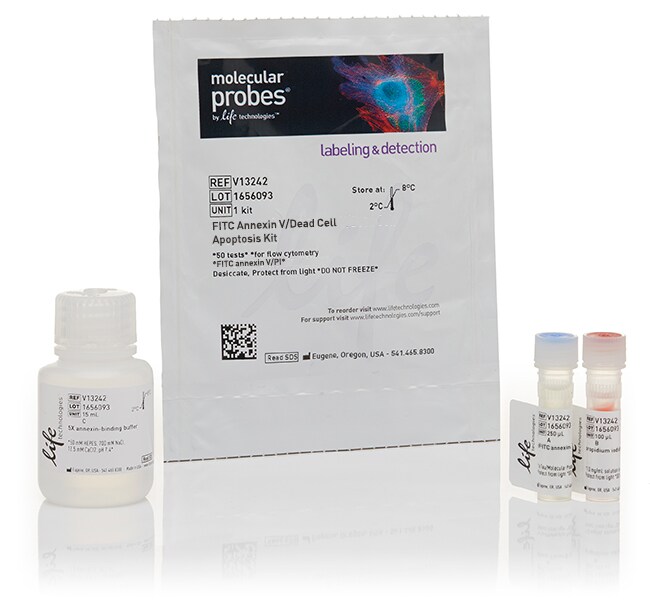
Catalog number V13242
Price (CNY)
2,820.00
飞享价
Ends: 31-Dec-2026
3,747.00Save 927.00 (25%)
1 kit
Quantity:
1 Kit
Label or Dye:
FITC, Propidium Iodide
Price (CNY)
2,820.00
飞享价
Ends: 31-Dec-2026
3,747.00Save 927.00 (25%)
1 kit
Easily differentiate live, dead, and apoptotic cells during flow cytometry with our Dead Cell Apoptosis kits with Annexin V conjugates including Alexa Fluor 488, FITC, propidium iodide, PE, APC, and SYTOX Green. These conjugates distinguish live, dead, or apoptotic cells via different dyes, which is vital for confirming cell viability and apoptosis using multi-parametric studies.
There are several advantages to using Dead Cell Apoptosis kits with Annexin V for flow cytometry in order to assay cell viability:
Superior brightness
Unlike other Annexin V kits that have lower protein concentrations or purification levels, the Annexin V Alexa Fluor 488 conjugate is optimized for flow cytometry to provide the largest separation between apoptotic and live cells. The Alexa Fluor 488 dye is a superior green-fluorescent dye with a spectrum similar to fluorescein (FITC).
High binding efficiency
Annexin V conjugates are made from a highly purified cys-annexin, resulting in higher binding efficiency and extremely accurate characterization of the apoptotic process.
Multi-parametric
Many publications require a minimum of two different ways to identify apoptotic cells. This multi-parametric kit detects phosphatidyl serine (PS) on the cytoplasmic surface of the cell membrane, while membrane integrity is determined via propidium iodide.
Dead Cell Apoptosis Kit with Annexin V Alexa Fluor 488 & PI
The Dead Cell Apoptosis Kit with Annexin V Alexa Fluor 488 & Propidium Iodide (PI) (V13241, V13245) is used in flow cytometry to measure early apoptosis by detecting phosphatidyl serine (PS) expression and membrane permeability. When cells are stained with Annexin V and propidium iodide, apoptotic cells expressing PS show green fluorescence, which can be detected in the FITC channel, and low red fluorescence. Dead or necrotic cells show bright red fluorescence and no green fluorescence, while live cells show no green or red fluorescence.
Dead Cell Apoptosis Kit with Annexin V PE and SYTOX Green
The Dead Cell Apoptosis Kit with Annexin V PE and SYTOX Green detects PS externalization in apoptotic cells during flow cytometry using recombinant annexin V conjugated to the orange fluorescent phycobiliprotein R-PE, while dead cells are detected with SYTOX Green nucleic acid stain. After treatment with both probes, apoptotic cells show orange fluorescence, dead cells show green fluorescence, and live cells show little or no fluorescence. These populations can easily be distinguished in the 530/30 nm and 585/42 nm bandpass filters with a 488 nm laser flow cytometer.
Dead Cell Apoptosis Kit with Annexin V APC and SYTOX Green
The Dead Cell Apoptosis Kit with Annexin V APC and SYTOX Green detects PS externalization in apoptotic cells during flow cytometry using recombinant annexin V conjugated to red laser-excited allophycocyanin, and dead cells using SYTOX Green nucleic acid stain. Apoptotic cells are detected by annexin V binding to externalized PS. Live cells show little or no fluorescence, apoptotic cells show red fluorescence and very little green fluorescence, and late apoptotic cells show a higher level of red and orange fluorescence. These populations can easily be distinguished using a flow cytometer that has both 488 nm and 633 nm excitation sources (an argon-ion laser and a HeNe laser).
The Dead Cell Apoptosis Kit with Annexin V FITC and PI
The Dead Cell Apoptosis Kit with Annexin V FITC and PI detects PS externalization in apoptotic cells using recombinant annexin V conjugated to green-fluorescent FITC dye and dead cells using PI. Necrotic cells that stain with PI show red fluorescence. After treatment with both probes, apoptotic cells show green fluorescence, dead cells show red and green fluorescence, and live cells show little or no fluorescence.
For Research Use Only. Not for use in diagnostic procedures.
Specifications
DescriptionDead Cell Apoptosis Kit with Annexin V FITC and PI, for flow cytometry
Excitation/Emission494, 535/518, 617
Flow Cytometer Laser Lines488
For Use With (Equipment)Flow Cytometer
Label or DyeFITC, Propidium Iodide
No. of Reactions50
Product TypeApoptosis Detection Kit
Quantity1 Kit
Shipping ConditionWet Ice
Unit Size1 kit
Contents & Storage
Contains 1 vial of annexin V, FITC conjugate (250 μL ), 1 vial of propidium iodide (PI, 100 μL), and 1 bottle of annexin binding buffer (5X solution, 15 mL). Store in refrigerator (2°C to 8°C) and protect from light.
Frequently asked questions (FAQs)
I want to study apoptosis using an Annexin V conjugate, but with adherent cells via microscopy instead of flow cytometry. Can this be done?
When using Dead Cell Apoptosis Kits with Annexin V for Flow Cytometry, what does a PI+ only population (hence Annexin V negative) correspond to?
I trypsinized my adherent cells and labeled with annexin V, and now my flow data is showing a high percentage of apoptotic cells even for control, untreated cells. What is the problem?
Can I detect annexin V staining in an imaging assay?
When should I stain adherent cells with annexin V for flow cytometric analysis? Before or after I trypsinize them?
Citations & References (34)
Citations & References
Abstract
Phenotypic and functional effects of heat shock protein 90 inhibition on dendritic cell.
Journal:Journal of immunology (Baltimore, Md. : 1950)
PubMed ID:17548610
Chitin is a size-dependent regulator of macrophage TNF and IL-10 production.
Journal:J Immunol
PubMed ID:19265136
'Chitin is a ubiquitous polysaccharide in fungi, insects, and parasites. We hypothesized that chitin is a size-dependent regulator of innate immunity. To test this hypothesis, we characterized the effects of chitins of different sizes on murine bronchoalveolar or peritoneal macrophages. In these studies, large chitin fragments were inert, while both
The ubiquitin-like protein FAT10 forms covalent conjugates and induces apoptosis.
Journal:J Biol Chem
PubMed ID:11445583
'FAT10 is a ubiquitin-like protein that is encoded in the major histocompatibility complex class I locus and is synergistically inducible with interferon-gamma and tumor necrosis factor alpha. The molecule consists of two ubiquitin-like domains in tandem arrangement and bears a conserved diglycine motif at its carboxyl terminus commonly used in
Apoptosis in tumour cells photosensitized with Rose Bengal acetate is induced by multiple organelle photodamage.
Journal:Histochem Cell Biol
PubMed ID:17849139
'Rose Bengal (RB) is a very efficient photosensitizer which undergoes inactivation of its photophysical and photochemical properties upon addition of a quencher group-i.e. acetate-to the xanthene rings. The resulting RB acetate (RB-Ac) derivative behaves as a fluorogenic substrate: it easily enters the cells where the native photoactive molecule is restored
Thiol/disulfide exchange is required for membrane fusion directed by the Newcastle disease virus fusion protein.
Journal:J Virol
PubMed ID:17151113
'Newcastle disease virus (NDV), an avian paramyxovirus, initiates infection with attachment of the viral hemagglutinin-neuraminidase (HN) protein to sialic acid-containing receptors, followed by fusion of viral and cell membranes, which is mediated by the fusion (F) protein. Like all class 1 viral fusion proteins, the paramyxovirus F protein is thought