Search
Invitrogen
CD326 (EpCAM) Monoclonal Antibody (G8.8), Brilliant Violet™ 711, eBioscience™
{{$productOrderCtrl.translations['antibody.pdp.commerceCard.promotion.promotions']}}
{{$productOrderCtrl.translations['antibody.pdp.commerceCard.promotion.viewpromo']}}
{{$productOrderCtrl.translations['antibody.pdp.commerceCard.promotion.promocode']}}: {{promo.promoCode}} {{promo.promoTitle}} {{promo.promoDescription}}. {{$productOrderCtrl.translations['antibody.pdp.commerceCard.promotion.learnmore']}}
Additional Information:
{{banner.description}}
 Antibody in Flow Cytometry (Flow)")
图: 1 / 1
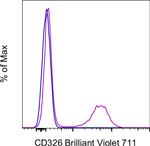
CD326 (EpCAM) Antibody (407-5791-82) in Flow
TE-71 cells were stained with 0.06 µg of Rat IgG2a kappa Isotype Control, Brilliant Violet 711 (Product # 407-4321-81) (blue histogram) or 0.06 µg of CD326 (EpCAM) Monoclonal Antibody, Brilliant Violet 711 (purple histogram). Viable cells were used for analysis, as determined by 7-AAD (Product # 00-6993-50).
Please note: We are reviewing Western blot images included in the antibody testing data in our catalog, including those provided by third parties. Unless expressly labeled or annotated as “raw-unedited”, Western blot images included in the antibody testing data in our catalog may have been edited, optimized or otherwise adjusted for presentation.
 Antibody (407-5791-82) in Flow")
产品信息
407-5791-82
产品规格
种属反应
Mouse
宿主/亚型
Rat
/ IgG2a, kappa
分类
Monoclonal
类型
Antibody
克隆号
G8.8
偶联物
Brilliant Violet™ 711
Brilliant Violet™ 711
Brilliant Violet™ 711
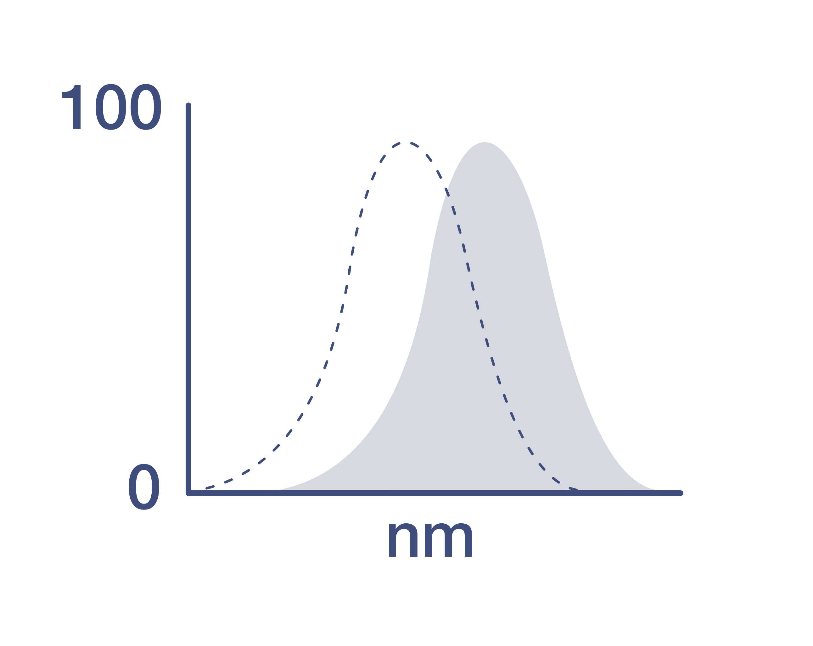
激发/发射光谱
406/711 nm
查看光谱
形式
Liquid
浓度
0.2 mg/mL
规格
100 µg
纯化类型
Affinity chromatography
保存液
PBS, pH 7.2, with BSA
内含物
0.09% sodium azide
保存条件
4°C, store in dark, DO NOT FREEZE!
运输条件
Ambient (domestic); Wet ice (international)
RRID
产品详细信息
Description
The G8.8 monoclonal antibody reacts with the 40 kDa mouse EpCAM (epithelial cellular adhesion molecule), also known as EGP40 (epithelial glycoprotein 40), 17-1A antigen, TACSTD1 (tumor-associated calcium signal transducer 1), and CD326. The immunogen used to generate the G8.8 antibody was the TE-71 thymic epithelial cell line. CD326 is expressed on the majority of epithelial cells, and is considered a pan-carcinoma antigen. CD326 mediates calcium-independent, homophilic, cell-cell adhesion and may function as a growth factor receptor. The antigen is being used as a target for immunotherapy treatment of human carcinomas. CD326 binds LAIR-1 (CD305) and LAIR-2 (CD306) to inhibit cellular activation and inflammation. This epithelial glycoprotein is now recognized as having an important role in tumor biology.
Applications Tested
This G8.8 antibody has been tested by flow cytometric analysis of TE-71 Cells. This may be used at less than or equal to 0.125 µg per test. A test is defined as the amount (µg) of antibody that will stain a cell sample in a final volume of 100 µL. Cell number should be determined empirically but can range from 10^5 to 10^8 cells/test. It is recommended that the antibody be carefully titrated for optimal performance in the assay of interest.
Blocking Buffers
When using two or more Super Bright, Brilliant Violet™, Brilliant Ultra Violet™, or other polymer dye-conjugated antibodies in a staining panel, it is recommended to use Super Bright Complete Staining Buffer (Product # SB-4401) or Brilliant Stain Buffer (Product # 00-4409-75) to minimize any non-specific polymer interactions. Please refer to the datasheet for Super Bright Staining Buffer or Brilliant Stain Buffer for more information.
Light sensitivity
This tandem dye is sensitive to photo-induced oxidation. Please protect this vial and stained samples from light.
Fixation
• Samples can be stored in IC Fixation Buffer (Product # 00-8222) (100 µL of cell sample + 100 µL of IC Fixation Buffer) or 1-step Fix/Lyse Solution (Product # 00-5333) for up to 3 days in the dark at 4°C with minimal impact on brightness and FRET efficiency/compensation.
• Some generalizations regarding fluorophore performance after fixation can be made, but clone specific performance should be determined empirically.
• Our internal testing suggests that Brilliant Violet™ 711 (BV711) is not compatible with methanol-based fixation.
Excitation: 407 nm; Emission: 713 nm; Laser: Violet Laser.
BRILLIANT ULTRA VIOLET™ is a trademark or registered trademark of Becton, Dickinson and Company or its affiliates, and is used under license. Powered by Sirigen™.
靶标信息
Ep-CAM (epithelial adhesion molecule, epithelial specific antigen, ESA) is a transmembrane glycoprotein expressed in the epithelium with a molecular weight of approximately 40 kDa, which functions as an epithelial cell adhesion molecule. Ep-CAM functions as a homotypic calcium-independent cell adhesion molecule, and has a direct impact on cell cycle, proliferation and metabolism of epithelial cells and fibroblasts due to its ability to rapidly induce the proto-oncogene c-myc and the cell cycle regulating genes cyclin A and E. Ep-CAM mediates Ca2+-independent homotypic interactions. Formation of Ep-CAM-mediated adhesions have a negative regulatory effect on adhesions mediated by classic cadherins, which may have strong effects on the differentiation and growth of epithelial cells. Ep-CAM overexpression was suggested to be associated with enhanced epithelial proliferation. Ep-CAM is highly expressed in human carcinomas, and is a marker for tumors of epithelial lineage. Ep-CAM is expressed on baso-lateral cell surface in most simple epithelia and many carcinoma types. Also, Ep-CAM reportedly distinguishes adenocarcinomas from pleural mesotheliomas.
仅用于科研。不用于诊断过程。未经明确授权不得转售。
How to use the Panel Builder
Watch the video to learn how to use the Invitrogen Flow Cytometry Panel Builder to build your next flow cytometry panel in 5 easy steps.
篇参考文献 (0)
您是否在文献中引用过该产品?请点击下方按钮邮件告知我们。
生物信息学
蛋白别名: CD326; EGP314; Ep-CAM; Epithelial cell adhesion molecule; Epithelial glycoprotein 314; hEGP314; lymphocyte antigen 74; mEGP314; panepithelial glycoprotein 314; Protein 289A; Trop-1 protein; Tumor-associated calcium signal transducer 1
基因别名: CD326; EGP; EGP-2; Egp314; Ep-CAM; Epcam; EpCAM1; GA733-2; gp40; Ly74; Tacsd1; Tacstd1; TROP1
UniProt ID: (Mouse) Q99JW5
Entrez Gene ID: (Mouse) 17075
positive regulation of cell proliferation
signal transduction involved in regulation of gene expression
negative regulation of apoptotic process
positive regulation of transcription from RNA polymerase II promoter
stem cell differentiation
cell-cell adhesion
negative regulation of cell-cell adhesion mediated by cadherin
positive regulation of cell motility
positive regulation of stem cell proliferation
Disclaimer

Clicking the images or links will redirect you to a website hosted by BenchSci that provides third-party scientific content. Neither the content nor the BenchSci technology and processes for selection have been evaluated by us; we are providing them as-is and without warranty of any kind, including for use or application of the Thermo Fisher Scientific products presented.

