Search
Invitrogen
Phospho-AKT1 (Ser473) Monoclonal Antibody (SDRNR), eFluor™ 450, eBioscience™
{{$productOrderCtrl.translations['antibody.pdp.commerceCard.promotion.promotions']}}
{{$productOrderCtrl.translations['antibody.pdp.commerceCard.promotion.viewpromo']}}
{{$productOrderCtrl.translations['antibody.pdp.commerceCard.promotion.promocode']}}: {{promo.promoCode}} {{promo.promoTitle}} {{promo.promoDescription}}. {{$productOrderCtrl.translations['antibody.pdp.commerceCard.promotion.learnmore']}}
Additional Information:
{{banner.description}}
 Antibody in Flow Cytometry (Flow)")
图: 1 / 11
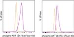
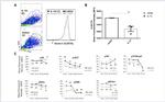
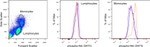
Phospho-AKT1 (Ser473) Antibody (48-9715-42) in Flow
Normal human peripheral blood cells were unstimulated (orange histogram) or were stimulated with Anti-Human CD3 and CD28 Functional Grade Purifieds (Product # 16-0037-81 and Product # 16-0289-81) (purple histogram). The cells were then intracellularly stained with Anti-Human/Mouse phospho-AKT (S473) eFluor® 450 and Anti-Human CD3 PerCP-Cyanine5-5 (Product # 45-0036-42) (left) or Anti-Human CD19 PE (Product ... View More
Please note: We are reviewing Western blot images included in the antibody testing data in our catalog, including those provided by third parties. Unless expressly labeled or annotated as “raw-unedited”, Western blot images included in the antibody testing data in our catalog may have been edited, optimized or otherwise adjusted for presentation.
 Antibody (48-9715-42) in Flow")
 Antibody (48-9715-42) in Flow")
 Antibody (48-9715-42) in Flow")
 Antibody (48-9715-42) in Flow")
 Antibody (48-9715-42) in Flow")
 Antibody (48-9715-42) in Flow")
 Antibody (48-9715-42) in Flow")
 Antibody (48-9715-42) in Flow")
 Antibody (48-9715-42) in Flow")
 Antibody (48-9715-42) in Flow")
 Antibody (48-9715-42)")
产品信息
48-9715-42
应用
建议稀释比
已发表文章
产品规格
种属反应
Human,
Mouse
已发表种属
Human,
Mouse
宿主/亚型
Mouse
/ IgG2a, kappa
分类
Monoclonal
类型
Antibody
克隆号
SDRNR
偶联物
eFluor™ 450
eFluor™ 450
eFluor™ 450
激发/发射光谱
405/445 nm
查看光谱
形式
Liquid
浓度
5 µL/Test
纯化类型
Affinity chromatography
保存液
PBS, pH 7.2, with BSA
内含物
0.09% sodium azide
保存条件
4°C, store in dark, DO NOT FREEZE!
运输条件
Ambient (domestic); Wet ice (international)
RRID
产品详细信息
Description: This SDRNR monoclonal antibody recognizes human and mouse AKT (also known as Protein Kinase B (PKB)) when phosphorylated on S473. AKT is a serine/threonine protein kinase that plays a key role in multiple cellular processes including metabolism, proliferation, apoptosis/survival, and migration. There are three homologous isoforms of AKT: AKT1, AKT2, and AKT3. AKT is activated by binding of its pleckstrin homology (PH) domain to membrane phospholipids and by phosphorylation. Phosphorylation of AKT at T308 by PDK1 and at S473 is required for full activation of this kinase. AKT promotes cell survival by inhibiting apoptosis via phosphorylation and inactivation of several targets including Bad, Foxo1, c-Raf, and caspase-9. Deregulation of AKT has been implicated as a major contributing factor in many types of cancer. AKT is negatively regulated by the phosphatase PTEN as well as by the chemical inhibitor LY294002. Specificity of this SDRNR clone was determined by ELISA, flow cytometry, and western blotting.
Applications Reported:This SDRNR antibody has been reported for use in intracellular staining followed by flow cytometric analysis.
Applications Tested: This SDRNR antibody has been pre-titrated and tested by intracellular staining followed by flow cytometric analysis of normal human peripheral blood mononuclear cells. This can be used at 5 µL (0.5 µg) per test. A test is defined as the amount (µg) of antibody that will stain a cell sample in a final volume of 100 µL. Cell number should be determined empirically but can range from 10^5 to 10^8 cells/test.
eFluor™ 450 is an alternative to Pacific Blue™. eFluor™ 450 emits at 445 nm and is excited with the Violet laser (405 nm). Please make sure that your instrument is capable of detecting this fluorochrome.
Staining Protocol: All protocols work well for this monoclonal antibody. Use of Protocol A: Two-step protocol: intracellular (cytoplasmic) proteins allows for the greatest flexibility for detection of surface and intracellular (cytoplasmic) proteins. Use of Protocol B: One-step protocol: intracellular (nuclear) proteins is recommended for staining of transcription factors in conjunction with surface and phosphorylated intracellular (cytoplasmic) proteins. Protocol C: Two-step protocol: Fixation/Methanol allows for the greatest discrimination of phospho-specific signaling between unstimulated and stimulated samples, but with limitations on the ability to stain specific surface proteins (refer to "Clone Performance Following Fixation/Permeabilization" located in the BestProtocols Section under the Resources tab online). All Protocols can be found in the Flow Cytometry Protocols: "Staining Intracellular Antigens for Flow Cytometry Protocol" located in the BestProtocols® Section under the Resources tab online.
Excitation: 405 nm; Emission: 445 nm; Laser: Violet Laser.
Filtration: 0.2 µm post-manufacturing filtered.
靶标信息
AKT1 (PKB alpha) is a serine/threonine kinase that regulates cell survival. The activated enzyme inhibits apoptosis and stimulates cell cycle progression by phosphorylating numerous targets in various cell types, including cancer cells. This protein kinase is activated by insulin, PI3K, IGF1 and various other growth and survival factors. Akt promotes cell survival by inhibiting apoptosis through phosphorylation and inactivation of several targets, including forkhead transcription factors, and caspase-9. The AKT pathway is a major target for cancer drug discovery.
仅用于科研。不用于诊断过程。未经明确授权不得转售。
How to use the Panel Builder
Watch the video to learn how to use the Invitrogen Flow Cytometry Panel Builder to build your next flow cytometry panel in 5 easy steps.
生物信息学
蛋白别名: AKT; AKT1 kinase; AKT1M; Akt1m protein; C-AKT; CAKT; PKB; PKB ALPHA; PKB beta; PKB gamma; PKB-ALPHA; PKBG; PRKBA; Protein kinase B; PROTEIN KINASE B ALPHA; protein kinase B-alpha; PROTO-ONCOGENE C-AKT; RAC; rac protein kinase alpha; RAC-ALPHA; RAC-ALPHA SERINE/THREONINE-PROTEIN KINASE; RAC-PK-ALPHA; RAC-PK-beta; RAC-PK-gamma; related to A and C kinases; serine-threonine protein kinase; Thymoma viral proto-oncogene; unnamed protein product; V-AKT MURINE THYMOMA VIRAL ONCOGENE HOMOLOG 1; V-AKT MURINE THYMOMA VIRAL ONCOGENE-LIKE PROTEIN 1
基因别名: AKT; AKT1; PKB; PKB-ALPHA; PKB/Akt; PKBalpha; PRKBA; RAC; RAC-ALPHA
UniProt ID: (Mouse) P31750
Entrez Gene ID: (Human) 207, (Mouse) 11651
protein kinase activity
protein serine/threonine kinase activity
protein serine/threonine/tyrosine kinase activity
protein binding
calmodulin binding
ATP binding
phosphatidylinositol-3,4,5-trisphosphate binding
kinase activity
enzyme binding
kinase binding
protein kinase binding
nitric-oxide synthase regulator activity
protein serine/threonine kinase inhibitor activity
identical protein binding
protein homodimerization activity
phosphatidylinositol-3,4-bisphosphate binding
14-3-3 protein binding
potassium channel activator activity
protein serine kinase activity
TORC2 complex binding
nucleotide binding
protein kinase C binding
transferase activity
transferase activity, transferring phosphorus-containing groups
protein phosphatase 2A binding
non-receptor serine/threonine protein kinase
osteoblast differentiation
positive regulation of endothelial cell proliferation
cell migration involved in sprouting angiogenesis
complement receptor mediated signaling pathway
sphingosine-1-phosphate signaling pathway
regulation of glycogen biosynthetic process
protein phosphorylation
nitric oxide biosynthetic process
activation-induced cell death of T cells
response to oxidative stress
signal transduction
epidermal growth factor receptor signaling pathway
G-protein coupled receptor signaling pathway
cell proliferation
insulin receptor signaling pathway
response to heat
response to hormone
negative regulation of autophagy
positive regulation of endothelial cell migration
positive regulation of gene expression
negative regulation of gene expression
negative regulation of plasma membrane long-chain fatty acid transport
fibroblast migration
positive regulation of glucose metabolic process
regulation of neuron projection development
negative regulation of macroautophagy
peptidyl-serine phosphorylation
peptidyl-threonine phosphorylation
cytokine-mediated signaling pathway
cell differentiation
positive regulation of cell growth
regulation of cell migration
positive regulation of cell migration
T cell costimulation
negative regulation of protein ubiquitination
TOR signaling
negative regulation of fatty acid beta-oxidation
positive regulation of endodeoxyribonuclease activity
positive regulation of proteasomal ubiquitin-dependent protein catabolic process
cellular response to insulin stimulus
positive regulation of peptidyl-serine phosphorylation
cellular response to stress
response to fluid shear stress
intracellular signal transduction
interleukin-18-mediated signaling pathway
positive regulation of protein import into nucleus
regulation of apoptotic process
negative regulation of apoptotic process
anoikis
regulation of mRNA stability
protein kinase B signaling
positive regulation of blood vessel endothelial cell migration
positive regulation of nitric oxide biosynthetic process
positive regulation of fat cell differentiation
positive regulation of glycogen biosynthetic process
negative regulation of Notch signaling pathway
negative regulation of proteolysis
positive regulation of transcription from RNA polymerase II promoter
nitric oxide metabolic process
positive regulation of glucose import
positive regulation of lipid biosynthetic process
insulin-like growth factor receptor signaling pathway
behavioral response to pain
positive regulation of smooth muscle cell proliferation
positive regulation of protein metabolic process
positive regulation of protein kinase B signaling
excitatory postsynaptic potential
response to growth hormone
mammary gland epithelial cell differentiation
response to UV-A
protein localization to mitochondrion
response to growth factor
cellular response to epidermal growth factor stimulus
maintenance of protein location in mitochondrion
cellular response to rapamycin
negative regulation of release of cytochrome c from mitochondria
vascular endothelial cell response to laminar fluid shear stress
regulation of postsynapse organization
regulation of tRNA methylation
cellular response to oxidised low-density lipoprotein particle stimulus
negative regulation of protein localization to lysosome
negative regulation of cGAS/STING signaling pathway
beta-arrestin-dependent dopamine receptor signaling pathway
positive regulation of G1/S transition of mitotic cell cycle
positive regulation of protein localization to nucleus
regulation of signal transduction by p53 class mediator
negative regulation of cilium assembly
negative regulation of oxidative stress-induced intrinsic apoptotic signaling pathway
negative regulation of leukocyte cell-cell adhesion
positive regulation of protein localization to plasma membrane
negative regulation of protein maturation
negative regulation of hydrogen peroxide-induced neuron intrinsic apoptotic signaling pathway
negative regulation of PERK-mediated unfolded protein response
positive regulation of TORC1 signaling
positive regulation of TORC2 signaling
positive regulation of protein localization to endoplasmic reticulum
positive regulation of anaphase-promoting complex-dependent catabolic process
cellular response to nerve growth factor stimulus
response to insulin-like growth factor stimulus
positive regulation of protein localization to cell surface
regulation of type B pancreatic cell development
negative regulation of lymphocyte migration
negative regulation of extrinsic apoptotic signaling pathway in absence of ligand
negative regulation of intrinsic apoptotic signaling pathway
protein import into nucleus, translocation
maternal placenta development
positive regulation of protein phosphorylation
carbohydrate metabolic process
glycogen metabolic process
glycogen biosynthetic process
glucose metabolic process
translation
regulation of translation
negative regulation of protein kinase activity
transport
apoptotic process
inflammatory response
cytoskeleton organization
multicellular organism development
germ cell development
nervous system development
aging
apoptotic mitochondrial changes
carbohydrate transport
positive regulation of fibroblast migration
positive regulation of sodium ion transport
negative regulation of endopeptidase activity
glucose transport
phosphorylation
protein ubiquitination
cell projection organization
protein catabolic process
regulation of myelination
positive regulation of cyclin-dependent protein serine/threonine kinase activity involved in G1/S transition of mitotic cell cycle
lipopolysaccharide-mediated signaling pathway
response to food
positive regulation of cellular protein metabolic process
peripheral nervous system myelin maintenance
regulation of protein localization
cellular response to vascular endothelial growth factor stimulus
cellular response to decreased oxygen levels
glucose homeostasis
anagen
positive regulation of apoptotic process
negative regulation of cysteine-type endopeptidase activity involved in apoptotic process
negative regulation of cell size
positive regulation of vasoconstriction
negative regulation of JNK cascade
positive regulation of nitric-oxide synthase activity
positive regulation of sequence-specific DNA binding transcription factor activity
striated muscle cell differentiation
glycogen cell differentiation involved in embryonic placenta development
labyrinthine layer blood vessel development
cellular response to mechanical stimulus
cellular response to growth factor stimulus
cellular response to prostaglandin E stimulus
cellular response to organic cyclic compound
cellular response to hypoxia
positive regulation of establishment of protein localization to plasma membrane
cellular response to granulocyte macrophage colony-stimulating factor stimulus
execution phase of apoptosis
cellular response to peptide
Disclaimer

Clicking the images or links will redirect you to a website hosted by BenchSci that provides third-party scientific content. Neither the content nor the BenchSci technology and processes for selection have been evaluated by us; we are providing them as-is and without warranty of any kind, including for use or application of the Thermo Fisher Scientific products presented.

