Search
Additional Information:
{{banner.description}}
")
FIGURE: 1 / 4
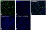
CD4 Antibody (740028TP488) in ICC/IF
Immunofluorescent analysis of CD4 was performed using CD4 transfected HEK-293E cells. The cells were fixed with 4% paraformaldehyde for 10 minutes, permeabilized with 0.1% Triton X-100 for 15 minutes and blocked with 2% BSA for 1 hour at room temperature. The cells were stained with CD4 Recombinant Rat Monoclonal Antibody (4SM95), Alexa Fluor™ Plus 488 (Product # 740028TP488, 1:800). Panel a) shows representative images of cells that were stained for detection and localization of CD4. Panel b) shows representative images of cells stained f... View More
Please note: We are reviewing Western blot images included in the antibody testing data in our catalog, including those provided by third parties. Unless expressly labeled or annotated as “raw-unedited”, Western blot images included in the antibody testing data in our catalog may have been edited, optimized or otherwise adjusted for presentation.
 in ICC/IF")
 in IHC (P)")
 in Flow")
")
Product Details
740028TP488
Applications
Tested Dilution
Publications
Product Specifications
Species Reactivity
Human,
Mouse
Host/Isotype
Rat
/ IgG1, kappa
Expression System
Expi293
Class
Recombinant Monoclonal
Type
Antibody
Clone
4SM95
Conjugate
Alexa Fluor™ Plus 488
Alexa Fluor™ Plus 488
Alexa Fluor™ Plus 488
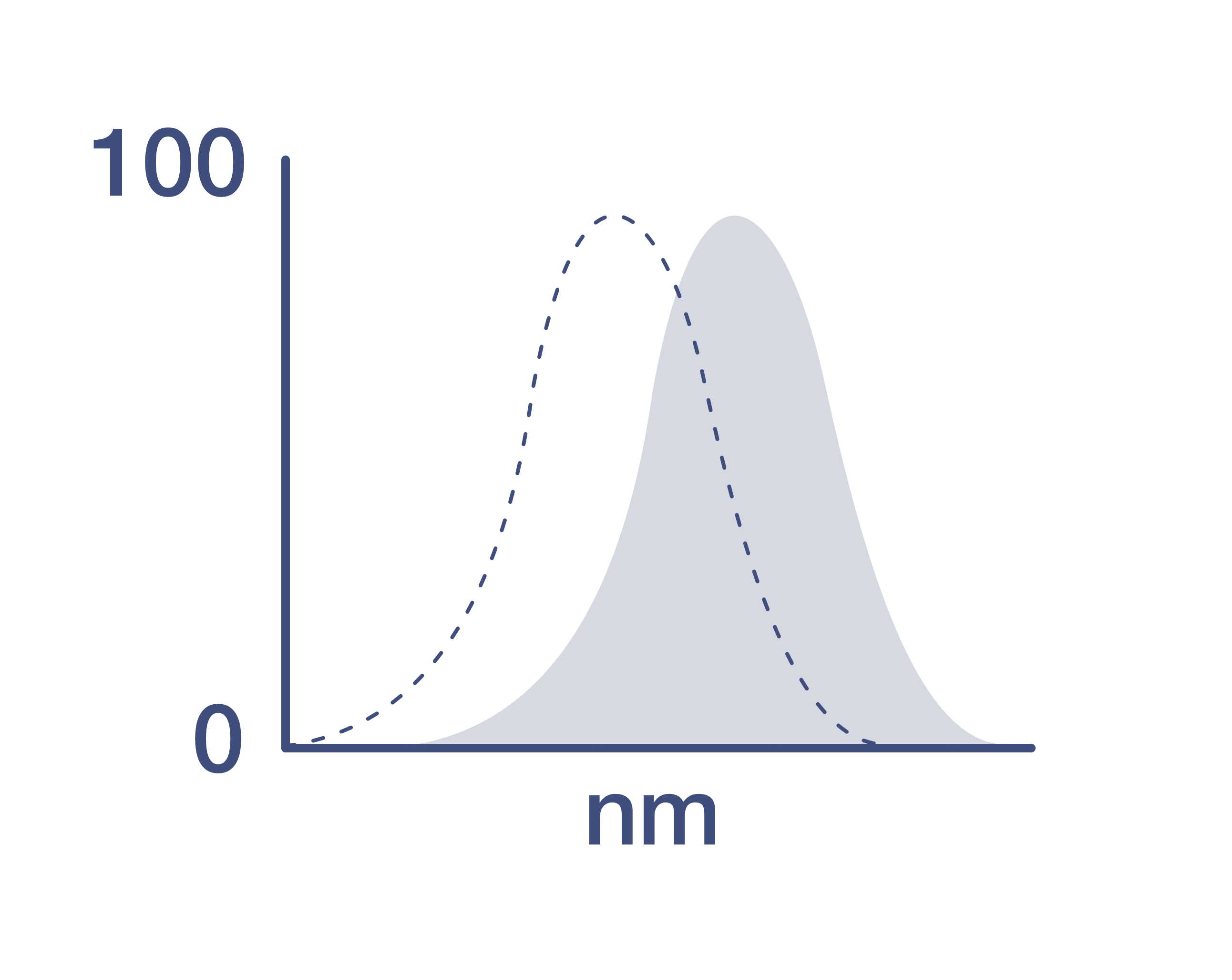
Excitation/Emission Max
493/518 nm
View spectra
Form
Liquid
Concentration
1.0 mg/mL
Amount
50 µg
Purification
Affinity chromatography
Storage buffer
proprietary buffer, pH 6.8
Contains
0.008% Bromonitrodioxane, 0.008% Methylisothiazolone
Storage conditions
4°C, store in dark, DO NOT FREEZE!
Shipping conditions
Wet ice
RRID
Product Specific Information
Alexa Fluor™ Plus recombinant rat antibodies are conjugated using new, proprietary dye chemistry so you can generate stunning data. Alexa Fluor™ Plus antibodies represent an advancement in fluorescent conjugate technology. Alexa Fluor™ Plus antibodies provide brighter signal compared to leading Alexa Fluor™ antibodies, providing you with better signal-to-noise for your critical experiments. These antibodies show better specificity and lot-to-lot consistency as these are recombinant antibodies, generated by cloning specific genes for the desired antibodies into an expression vector and expressed in vitro.
Using conjugate solutions: Centrifuge the protein conjugate solution briefly in a microcentrifuge before use; add only the supernatant to the experiment. This step will help eliminate any protein aggregates that may have formed during storage, thereby reducing nonspecific background staining.
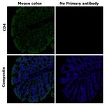
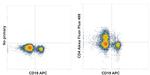
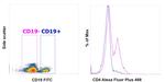
Applications Tested: This 4SM95 antibody has been tested by immunohistochemistry of colon and immunocytochemistry of CD4 transfected HEK-293E cells and flow cytometric analysis of human PBMC. This may be used for immunohistochemistry at 10 µg/mL and for immunocytochemistry at 1.25 µg/mL and for flow cytometry at less than or equal to 1 µg per test. A test is defined as the amount (µg) of antibody that will stain a cell sample in a final volume of 100 µL. Cell number should be determined empirically but can range from 10^5 to 10^8 cells/test. It is recommended that the antibody be carefully titrated for optimal performance in the assay of interest.
Excitation: 494 nm; Emission: 519 nm; Laser: Blue Laser
Filtration: 0.2 µm post-manufacturing filtered.
Target Information
The CD4 antigen is involved in the recognition of MHC class II molecules and is a co-receptor for HIV. CD4 is primarily expressed in a subset of T-lymphocytes, also referred to as T helper cells, but may also be expressed by other cells in the immune system, such as monocytes, macrophages, and dendritic cells. At the tissue level, CD4 expression may be detected in thymus, lymph nodes, tonsils, and spleen, and also in specific regions of the brain, gut, and other non-lymphoid tissues. CD4 functions to initiate or augment the early phase of T-cell activation through its association with the T-cell receptor complex and protein tyrosine kinase, Lck. It may also function as an important mediator of direct neuronal damage in infectious and immune-mediated diseases of the central nervous system. Multiple alternatively spliced transcripts have been identified in this gene [RefSeq, July 2017].
For Research Use Only. Not for use in diagnostic procedures. Not for resale without express authorization.
References (0)
Have you cited this product in a publication?
Let us know so we can reference it here.
Bioinformatics
Protein Aliases: CD4; CD4 antigen p55; cd4a; fCD4; Leu-3; T-cell differentiation antigen L3T4; T-cell surface antigen T4/Leu-3; T-cell surface glycoprotein CD4; T-cell surface glycoprotein CD4 precursor (T-cell surface antigen T4/Leu-3) (T-cell differentiation antigen L3T4)
Gene Aliases: CD4; L3T4; Ly-4
UniProt ID: (Mouse) P06332
Entrez Gene ID: (Mouse) 12504
cytokine production
immune system process
induction by virus of host cell-cell fusion
immune response
cell adhesion
cell surface receptor signaling pathway
T cell differentiation
maintenance of protein location in cell
T cell activation
T cell selection
protein palmitoleylation
positive regulation of protein kinase activity
positive regulation of peptidyl-tyrosine phosphorylation
defense response to Gram-negative bacterium
positive regulation of calcium-mediated signaling
regulation of T cell activation
positive regulation of T cell activation
Disclaimer

Clicking the images or links will redirect you to a website hosted by BenchSci that provides third-party scientific content. Neither the content nor the BenchSci technology and processes for selection have been evaluated by us; we are providing them as-is and without warranty of any kind, including for use or application of the Thermo Fisher Scientific products presented.

Performance Guarantee
If an Invitrogen™ antibody doesn't perform as described on our website or datasheet,we'll replace the product at no cost to you, or provide you with a credit for a future purchase.*
Learn more
We're here to help
Get expert recommendations for common problems or connect directly with an on staff expert for technical assistance related to applications, equipment and general product use.
Contact tech support