Search
Invitrogen
Arginase 1 Recombinant Rat Monoclonal Antibody (A1exF5), Alexa Fluor™ Plus 647
{{$productOrderCtrl.translations['antibody.pdp.commerceCard.promotion.promotions']}}
{{$productOrderCtrl.translations['antibody.pdp.commerceCard.promotion.promocode']}}: {{promo.promoCode}} {{promo.promoTitle}} {{promo.promoDescription}}. {{$productOrderCtrl.translations['antibody.pdp.commerceCard.promotion.learnmore']}}
{{$ctrl.translations['antibody.pdp.skuNotes.additionalInformation']}}:
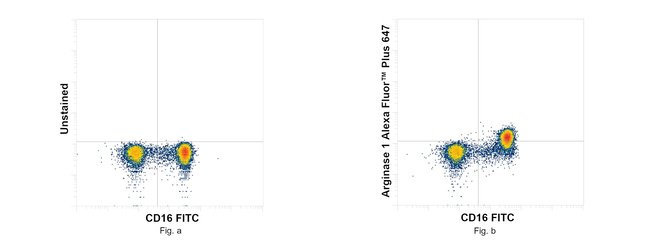
图: 1 / 3
Arginase 1 Antibody (750030TP647) in Flow
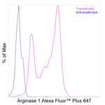
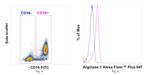
Flow cytometry analysis of Arginase 1 Recombinant Rat Monoclonal Antibody (A1exF5), Alexa Fluor™ Plus 647 (Product # 750030TP647) was performed using normal human lysed whole blood cells. Normal human lysed whole blood cells were fixed with eBioscience™ IC Fixation Buffer (Product # 00-8222-49), permeabilized, and stained with CD16 Monoclonal Antibody (eBioCB16 (CB16)), FITC, eBioscience™, (Product # 11-0168-42) and 0.5 µg of Arginase 1 Recombinant Rat Monoclonal Antibody (A1exF5), Alexa Fluor™ Plus 647 (Product # 750030TP647) (Fig. b) or ... View More
Please note: We are reviewing Western blot images included in the antibody testing data in our catalog, including those provided by third parties. Unless expressly labeled or annotated as “raw-unedited”, Western blot images included in the antibody testing data in our catalog may have been edited, optimized or otherwise adjusted for presentation.


产品信息
750030TP647
产品规格
种属反应
Human
宿主/亚型
Rat
/ IgG2a, kappa
Expression System
Expi293
分类
Recombinant Monoclonal
类型
Antibody
克隆号
A1exF5
偶联物
Alexa Fluor™ Plus 647
Alexa Fluor™ Plus 647
Alexa Fluor™ Plus 647
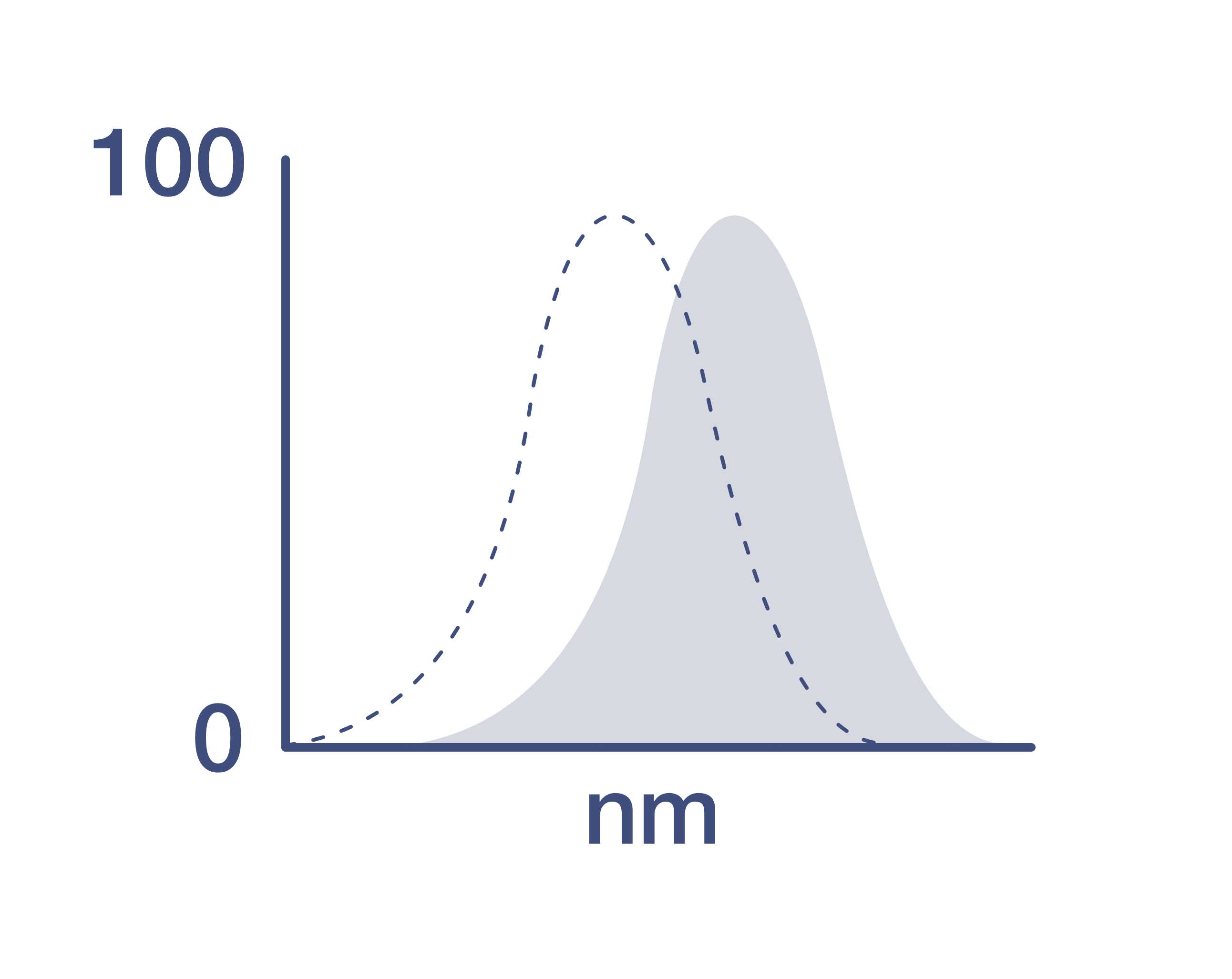
激发/发射光谱
658/675 nm
查看光谱
形式
Liquid
浓度
1 mg/mL
规格
50 µg
纯化类型
Affinity chromatography
保存液
proprietary buffer, pH 6.3-7.3
内含物
0.008% Bromonitrodioxane, 0.008% Methylisothiazolone
保存条件
4°C, store in dark, DO NOT FREEZE!
运输条件
Wet ice
产品详细信息
Alexa Fluor™ Plus recombinant antibodies are conjugated using new, proprietary dye chemistry so you can generate stunning data. Alexa Fluor™ Plus antibodies represent an advancement in fluorescent conjugate technology. Alexa Fluor™ Plus antibodies provide brighter signal compared to leading Alexa Fluor™ antibodies, providing you with better signal-to-noise for your critical experiments. These antibodies show better specificity and lot-to-lot consistency as these are recombinant antibodies, generated by cloning specific genes for the desired antibodies into an expression vector and expressed in vitro.
Using conjugate solutions: Centrifuge the protein conjugate solution briefly in a microcentrifuge before use; add only the supernatant to the experiment. This step will help eliminate any protein aggregates that may have formed during storage, thereby reducing nonspecific background staining.
It is recommended that the antibody be carefully titrated for optimal performance in the assay of interest.
Excitation: 658 nm; Emission: 675 nm; Laser: Red Laser
Filtration: 0.2 µm post-manufacturing filtered.
靶标信息
Arginase-1 (Arg1) is a 35 kDa enzyme converting L-arginine to urea and L-ornithine, which is the final step in the urea cycle. The resulting polyamines are important for cell proliferation and removal of toxins that arise from protein degradation. By degrading arginine, Arginase 1 deprives NO synthase of its substrate and down-regulates nitric oxide production. In both human and mouse, Arginase 1 is expressed in the liver, neutrophils, myeloid derived suppressor cells (MDSC) and neural stem cells. In human, expression in blood neutrophils but not in CCR3+ granulocytes has been reported. In mice, expression of Arginase 1 is one of the hallmarks of alternatively activated macrophages (M2a). Arginase-1 may be expressed in the myeloid cells infiltrating tumors, and is typically found in the majority of hepatocellular carcinomas. Defects in Arginase 1 are the cause of argininemia, an autosomal recessive disorder characterized by hyperammonemia.
仅用于科研。不用于诊断过程。未经明确授权不得转售。
篇参考文献 (0)
您是否在文献中引用过该产品?请点击下方按钮邮件告知我们。
生物信息学
蛋白别名: A-I; Arginase; arginase (EC 3.5.3.1); arginase 1 liver; arginase, liver; Arginase1; HGNC:663; Liver Arginase; liver-type arginase; Type 1 Arginase; type I arginase
UniProt ID: (Human) P05089
Entrez Gene ID: (Human) 383
urea cycle
arginine metabolic process
arginine catabolic process
regulation of cell proliferation
negative regulation of T cell proliferation
defense response to protozoan
negative regulation of activated T cell proliferation
negative regulation of interferon-gamma-mediated signaling pathway
neutrophil mediated killing of fungus
negative regulation of T-helper 2 cell cytokine production
Disclaimer

Clicking the images or links will redirect you to a website hosted by BenchSci that provides third-party scientific content. Neither the content nor the BenchSci technology and processes for selection have been evaluated by us; we are providing them as-is and without warranty of any kind, including for use or application of the Thermo Fisher Scientific products presented.

