Search
Additional Information:
{{banner.description}}
 (IHC (P))")
FIGURE: 1 / 5
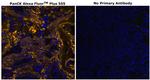
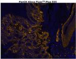
Cytokeratin Pan Type I/II Antibody (754-9003-82) in IHC (P)
Immunohistochemical analysis of Pan-CK was performed on formalin-fixed paraffin-embedded human tonsil tissue. To expose the target protein, HIER was performed on de-paraffinized sections using BOND Epitope Retrieval Solution 2 (pH 9) for 10 minutes, followed by a 5-min cool down and a 5-min wash with ddH2O. Tissues were permeabilized with 0.1% Triton X-100 in 1X PBS for 30 mins and blocked with 3% BSA/5% no... View More

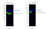
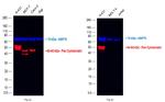
Please note: We are reviewing Western blot images included in the antibody testing data in our catalog, including those provided by third parties. Unless expressly labeled or annotated as “raw-unedited”, Western blot images included in the antibody testing data in our catalog may have been edited, optimized or otherwise adjusted for presentation.
 in IHC (P)")
 in IHC (P)")
 in IHC (P)")
")
")
Product Details
754-9003-82
Applications
Tested Dilution
Publications
Product Specifications
Species Reactivity
Human
Host/Isotype
Mouse
/ IgG1
Class
Monoclonal
Type
Antibody
Clone
AE1/AE3
Immunogen
Human epidermal keratin
Conjugate
Alexa Fluor™ Plus 555
Alexa Fluor™ Plus 555
Alexa Fluor™ Plus 555
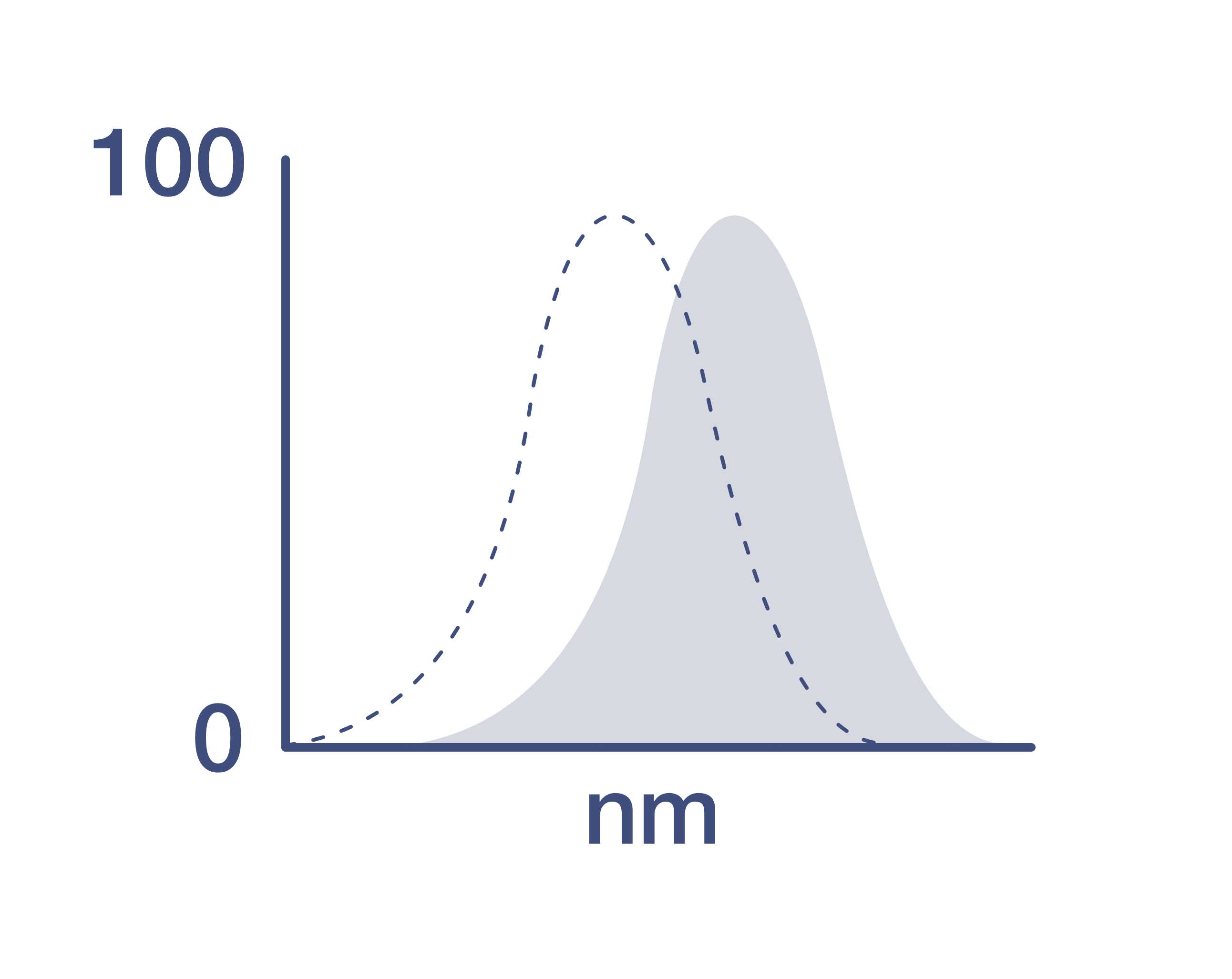
Excitation/Emission Max
558/572 nm
View spectra
Form
Liquid
Concentration
0.2 mg/mL
Amount
100 µg
Purification
Affinity chromatography
Storage buffer
PBS, pH 7.2, with 0.5% BSA, 10% proprietary stabilizer
Contains
0.05% sodium azide
Storage conditions
4°C, store in dark
Shipping conditions
Wet ice
RRID
Product Specific Information
Description: The monoclonal antibodies AE1 and AE3 recognize many of the acidic and basic cytokeratin family members. Cytokeratins are intermediate filament proteins comprising one component of the cytoskeleton. There are two large families of cytokeratins, acidic and basic, but all contain the same basic domains (i.e. an alpha-helical core with an N- and C-terminal domain). The proteins are expressed in epithelial cells, but are developmentally regulated. Many tumors also express these proteins and their expression can help identify the origin of a neoplasm.
The AE3 monoclonal antibody recognizes the 65 to 67 triplet, 64, 59, 58, 56, 54 and 52kD proteins also known as cytokeratin 1, 2, 3, 4, 5, 6, 7, and 8 while the AE1 antibody recognizes 56.5, 54', 50, 50', 48, and 40 kDa proteins (also known as CK10, 14, 15, 16 and 19). These antibodies can be used on a wide array of tissue samples from mouse, human, rat, primates (cynomolgus and rhesus), dog, cat, rabbit, and chicken.
Applications Reported: This AE1/AE3 antibody has been reported for use in immunohistochemical staining, immunocytochemistry, immunohistochemical staining of frozen tissue sections, and immunohistochemical staining of formalin-fixed paraffin embedded tissue sections.
Applications Tested: This AE1/AE3 antibody has been tested by immunohistochemistry of formalin-fixed paraffin embedded tissue using high pH antigen retrieval conditions and can be used at 4 µg/mL. It is recommended that the antibody be carefully titrated for optimal performance in the assay of interest.
Using conjugate solutions: Centrifuge the protein conjugate solution briefly in a microcentrifuge before use; add only the supernatant to the experiment. This step will help eliminate any protein aggregates that may have formed during storage, thereby reducing nonspecific background staining.
Target Information
Cytokeratin pan is part of a subfamily of intermediate filament proteins that are characterized by remarkable biochemical diversity, and represented in human epithelial tissues by at least 20 different polypeptides. Cytokeratins range in molecular weight between 40 kDa- 68 kDa, and an isoelectric pH between 4.9-7.8. The individual human cytokeratins are numbered 1 to 20. The various epithelia in the human body usually express cytokeratins which are not only characteristic of the type of epithelium, but also related to the degree of maturation or differentiation within an epithelium. Cytokeratin subtype expression patterns are used to an increasing extent in the distinction of different types of epithelial malignancies. The cytokeratin antibodies are not only of assistance in the differential diagnosis of tumors using immunohistochemistry on tissue sections, but are also a useful tool in cytopathology and flow cytometric assays. The composition of cytokeratin pairs vary with the epithelial cell type, stage of differentiation, cellular growth environment, and disease state. Many studies have shown the usefulness of keratins as markers in cancer research and tumor diagnosis.
For Research Use Only. Not for use in diagnostic procedures. Not for resale without express authorization.
References (0)
Have you cited this product in a publication?
Let us know so we can reference it here.
Bioinformatics
Protein Aliases: 39.1; 58 kDa cytokeratin; 65 kDa cytokeratin; 67 kDa cytokeratin; Cell proliferation-inducing gene 46 protein; CK-1; CK-10; CK-13; CK-14; CK-15; CK-16; CK-17; CK-18; CK-19; CK-1B; CK-2e; CK-2P; CK-3; CK-4; CK-5; CK-6A; CK-6D; CK-7; CK-8; Cytokeratin-1; Cytokeratin-10; Cytokeratin-13; Cytokeratin-14; Cytokeratin-15; Cytokeratin-16; Cytokeratin-17; Cytokeratin-18; Cytokeratin-19; Cytokeratin-1B; Cytokeratin-2e; Cytokeratin-2P; Cytokeratin-3; Cytokeratin-4; Cytokeratin-5; Cytokeratin-6A; Cytokeratin-6D; Cytokeratin-7; Cytokeratin-8; Epithelial keratin-2e; Hair alpha protein; HMWCK; K1; K10; K13; K14; K15; K16; K17; K18; K19; K1B; K2e; K3; K4; K5; K6A; K7; K76; K77; K8; Keratin 1B; Keratin type II cytoskeletal 5; Keratin, type I cytoskeletal 10; Keratin, type I cytoskeletal 13; Keratin, type I cytoskeletal 14; Keratin, type I cytoskeletal 15; Keratin, type I cytoskeletal 16; Keratin, type I cytoskeletal 17; Keratin, type I cytoskeletal 18; Keratin, type I cytoskeletal 19; Keratin, type II cytoskeletal 1; Keratin, type II cytoskeletal 1b; Keratin, type II cytoskeletal 2 epidermal; Keratin, type II cytoskeletal 2 oral; Keratin, type II cytoskeletal 3; Keratin, type II cytoskeletal 4; Keratin, type II cytoskeletal 5; Keratin, type II cytoskeletal 6A; Keratin, type II cytoskeletal 7; Keratin, type II cytoskeletal 8; Keratin-1; Keratin-10; Keratin-13; Keratin-14; Keratin-15; Keratin-16; Keratin-17; Keratin-18; Keratin-19; Keratin-2 epidermis; Keratin-2e; Keratin-3; Keratin-4; Keratin-5; Keratin-6A; Keratin-7; Keratin-76; Keratin-77; Keratin-8; KRT1B; Pan CK; pan Cytokeratin; pan keratin; pankeratin; Sarcolectin; type II cytoskeletal 1b; Type-II keratin Kb1; Type-II keratin Kb2; Type-II keratin Kb3; Type-II Keratin Kb39; Type-II keratin Kb4; Type-II keratin Kb5; Type-II keratin Kb6; Type-II keratin Kb7; Type-II keratin Kb8; Type-II keratin Kb9
Gene Aliases: CYK18; CYK4; CYK8; K6A; KPP; KRT1; KRT10; KRT13; KRT14; KRT15; KRT16; KRT16A; KRT17; KRT18; KRT19; KRT1B; KRT2; KRT2A; KRT2B; KRT2E; KRT2P; KRT3; KRT4; KRT5; KRT6A; KRT6D; KRT7; KRT76; KRT77; KRT8; KRTA; KRTB; PIG46; SCL
Disclaimer

Clicking the images or links will redirect you to a website hosted by BenchSci that provides third-party scientific content. Neither the content nor the BenchSci technology and processes for selection have been evaluated by us; we are providing them as-is and without warranty of any kind, including for use or application of the Thermo Fisher Scientific products presented.

Performance Guarantee
If an Invitrogen™ antibody doesn't perform as described on our website or datasheet,we'll replace the product at no cost to you, or provide you with a credit for a future purchase.*
Learn more
We're here to help
Get expert recommendations for common problems or connect directly with an on staff expert for technical assistance related to applications, equipment and general product use.
Contact tech support