Search
Invitrogen
CD63 Monoclonal Antibody (NVG-2), PE-Cyanine7, eBioscience™
{{$productOrderCtrl.translations['antibody.pdp.commerceCard.promotion.promotions']}}
{{$productOrderCtrl.translations['antibody.pdp.commerceCard.promotion.viewpromo']}}
{{$productOrderCtrl.translations['antibody.pdp.commerceCard.promotion.promocode']}}: {{promo.promoCode}} {{promo.promoTitle}} {{promo.promoDescription}}. {{$productOrderCtrl.translations['antibody.pdp.commerceCard.promotion.learnmore']}}
Additional Information:
{{banner.description}}
")
图: 1 / 2
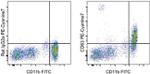
CD63 Antibody (25-0631-82) in Flow
Intracellular staining of mouse resident peritoneal exudate cells with Anti-Mouse CD11b FITC (Product # 11-0112-41) and 0.06 µg of Rat IgG2a K Isotype Control PE-Cyanine7 (Product # 25-4321-82) (left) or 0.06 µg of Anti-Mouse CD63 PE-Cyanine7 (right) using the Intracellular Fixation & Permeabilization Buffer Set (Product # 88-8824-00) and protocol. Total viable cells, as determined by Fixable Viability Dye eFluor® 506 (Product # 65-0866-14), were used for analysis.
 in Flow")
 in Flow")
产品信息
25-0631-82
应用
建议稀释比
已发表文章
产品规格
种属反应
Mouse
宿主/亚型
Rat
/ IgG2a, kappa
分类
Monoclonal
类型
Antibody
克隆号
NVG-2
偶联物
PE-Cyanine7
PE-Cyanine7
PE-Cyanine7
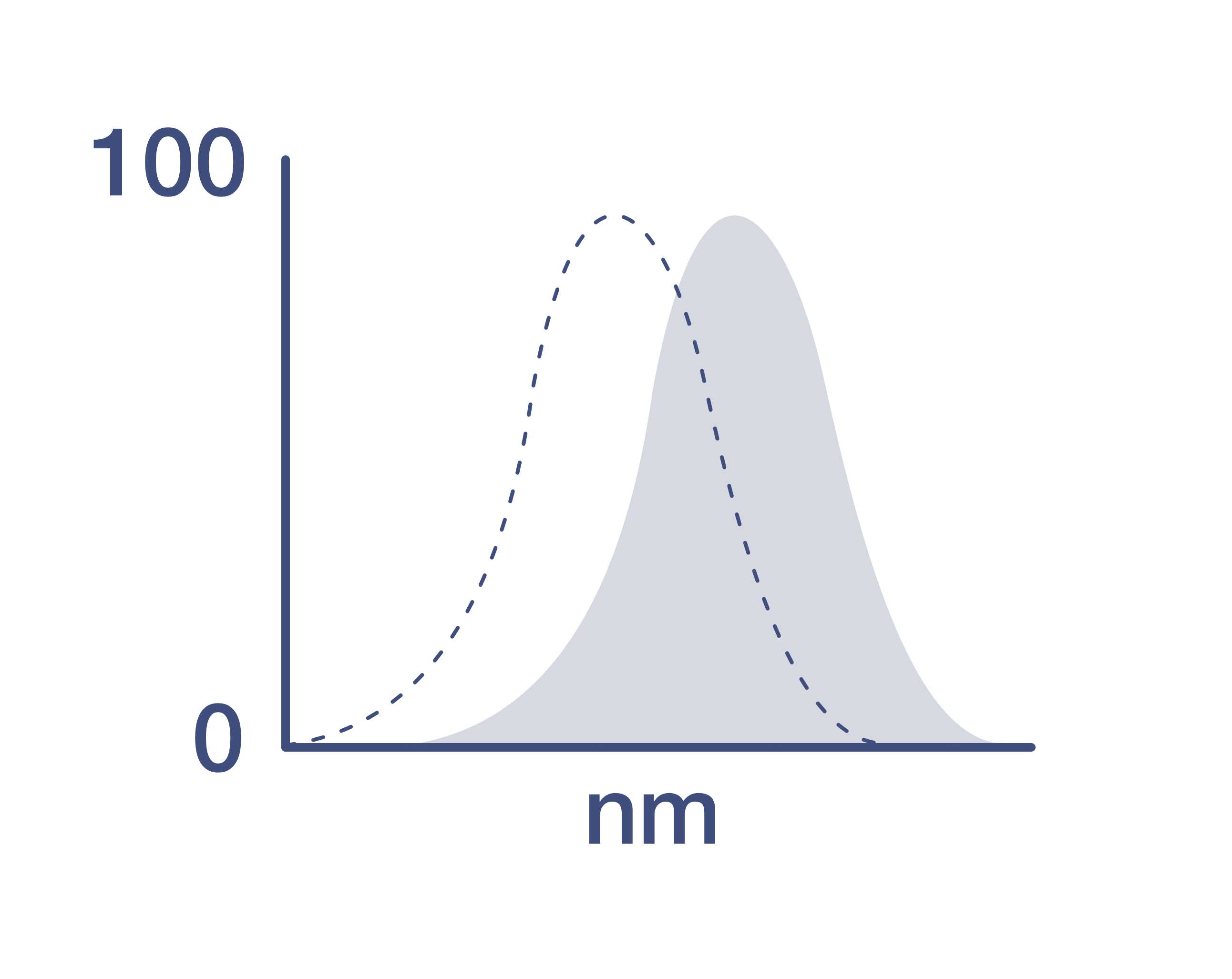
激发/发射光谱
569/780 nm
查看光谱
形式
Liquid
浓度
0.2 mg/mL
规格
100 µg
纯化类型
Affinity chromatography
保存液
PBS, pH 7.2
内含物
0.09% sodium azide
保存条件
4°C, store in dark, DO NOT FREEZE!
运输条件
Ambient (domestic); Wet ice (international)
RRID
产品详细信息
Description: The monoclonal antibody NVG-2 reacts with mouse CD63, also known as Lysosomal-Associated Membrane Protein 3 (LAMP-3) or tetraspanin 30 (TSPN30), a member of tetraspanin family of proteins characterized by four transmembrane domains. CD63 is expressed on a variety of cell types of hematopoietic lineage, e.g., granulocytes, B lymphocytes, platelets, as well as cells of non-hematopoietic origin. It can be found on the cell membrane, late endocytic vesicles, lysosomes, exosomes, and other specialized granules. On the cell surface, CD63 has been shown to interact with various proteins forming tetraspanin-enriched microdomains (TEM). Its high expression on the cell membrane may be indicative of cell activation, hence, CD63 is often used as an activation marker for basophils, platelets and other cells.
Applications Reported: This NVG-2 antibody has been reported for use in intracellular staining followed by flow cytometric analysis.
Applications Tested: This NVG-2 antibody has been tested by intracellular staining followed by flow cytometric analysis of mouse resident peritoneal exudate cells using the Intracellular Fixation & Permeabilization Buffer Set (Product # 88-8824-00) and protocol. Please refer to BestProtocols®: Protocol A: Two step protocol for (cytoplasmic) intracellular proteins located under the Resources Tab online. This can be used at less than or equal to 0.125 µg per test. A test is defined as the amount (µg) of antibody that will stain a cell sample in a final volume of 100 µL. Cell number should be determined empirically but can range from 10^5 to 10^8 cells/test. It is recommended that the antibody be carefully titrated for optimal performance in the assay of interest.
Light sensitivity: This tandem dye is sensitive to photo-induced oxidation. Please protect this vial and stained samples from light.
Fixation: Samples can be stored in IC Fixation Buffer (Product # 00-822-49) (100 µL of cell sample + 100 µL of IC Fixation Buffer) or 1-step Fix/Lyse Solution (Product # 00-5333-54) for up to 3 days in the dark at 4°C with minimal impact on brightness and FRET efficiency/compensation. Some generalizations regarding fluorophore performance after fixation can be made, but clone specific performance should be determined empirically.
Excitation: 488-561 nm; Emission: 775 nm; Laser: Blue Laser, Green Laser, Yellow-Green Laser.
Filtration: 0.2 µm post-manufacturing filtered.
靶标信息
CD63, also known as LAMP-3, is a glycoprotein belonging to the tetraspanin family, primarily located in late endosomes, lysosomes, and secretory vesicles across various cell types. It is expressed intracellularly in resting platelets and basophils, but upon activation, CD63 translocates to the plasma membrane, making it a widely used marker for basophil activation. Unlike basophils, mast cells can expose CD63 without activation. CD63 is expressed on activated platelets, monocytes, macrophages, and granulocytes, as well as endothelial cells, fibroblasts, and smooth muscle cells. It interacts with integrins (such as VLA-3 and VLA-6) and TIMP-1, playing roles in mediating allergic responses, phagocytosis, and cell migration. Additionally, CD63 is involved in the regulation of H/K-ATPase trafficking and ROMK1 channels, and serves as a T-cell costimulation molecule. CD63 expression can be used to predict prognosis in early-stage carcinomas and is identical to the melanoma-associated antigen ME491 and the platelet antigen PTLGP40. Diseases associated with CD63 dysfunction include melanoma and Hermansky-Pudlak Syndrome, highlighting its significance in both immune responses and disease processes.
仅用于科研。不用于诊断过程。未经明确授权不得转售。
How to use the Panel Builder
Watch the video to learn how to use the Invitrogen Flow Cytometry Panel Builder to build your next flow cytometry panel in 5 easy steps.
生物信息学
蛋白别名: CD 63; CD63; CD63 antigen; melanoma 1 antigen
基因别名: C75951; Cd63; ME491; Tspan30
UniProt ID: (Mouse) P41731
Entrez Gene ID: (Mouse) 12512
positive regulation of receptor internalization
transport
cell-matrix adhesion
negative regulation of epithelial cell migration
protein transport
cell migration
epithelial cell differentiation
cellular protein localization
endosome to melanosome transport
pigmentation
positive regulation of cell adhesion
positive regulation of endocytosis
pigment granule maturation
pigment cell differentiation
regulation of vascular endothelial growth factor signaling pathway
regulation of rubidium ion transport
positive regulation of integrin-mediated signaling pathway
Disclaimer

Clicking the images or links will redirect you to a website hosted by BenchSci that provides third-party scientific content. Neither the content nor the BenchSci technology and processes for selection have been evaluated by us; we are providing them as-is and without warranty of any kind, including for use or application of the Thermo Fisher Scientific products presented.

