Search
Invitrogen
CD7 Monoclonal Antibody (4H9), PerCP-eFluor™ 710, eBioscience™
{{$productOrderCtrl.translations['antibody.pdp.commerceCard.promotion.promotions']}}
{{$productOrderCtrl.translations['antibody.pdp.commerceCard.promotion.viewpromo']}}
{{$productOrderCtrl.translations['antibody.pdp.commerceCard.promotion.promocode']}}: {{promo.promoCode}} {{promo.promoTitle}} {{promo.promoDescription}}. {{$productOrderCtrl.translations['antibody.pdp.commerceCard.promotion.learnmore']}}
Additional Information:
{{banner.description}}
")
图: 1 / 1
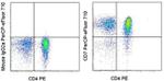
CD7 Antibody (46-0078-42) in Flow
Staining of normal human peripheral blood cells with Anti-Human CD4 PE (Product # 12-0048-42) and Mouse IgG2a K Isotype Control PerCP-eFluor® 710 (Product # 46-4724-82) (left) or Anti-Human CD7 PerCP-eFluor® 710 (right). Cells in the lymphocyte gate were used for analysis.
Please note: We are reviewing Western blot images included in the antibody testing data in our catalog, including those provided by third parties. Unless expressly labeled or annotated as “raw-unedited”, Western blot images included in the antibody testing data in our catalog may have been edited, optimized or otherwise adjusted for presentation.
 in Flow")
产品信息
46-0078-42
应用
建议稀释比
已发表文章
产品规格
种属反应
Human
已发表种属
Human
宿主/亚型
Mouse
/ IgG2a, kappa
分类
Monoclonal
类型
Antibody
克隆号
4H9
偶联物
PerCP-eFluor™ 710
PerCP-eFluor™ 710
PerCP-eFluor™ 710
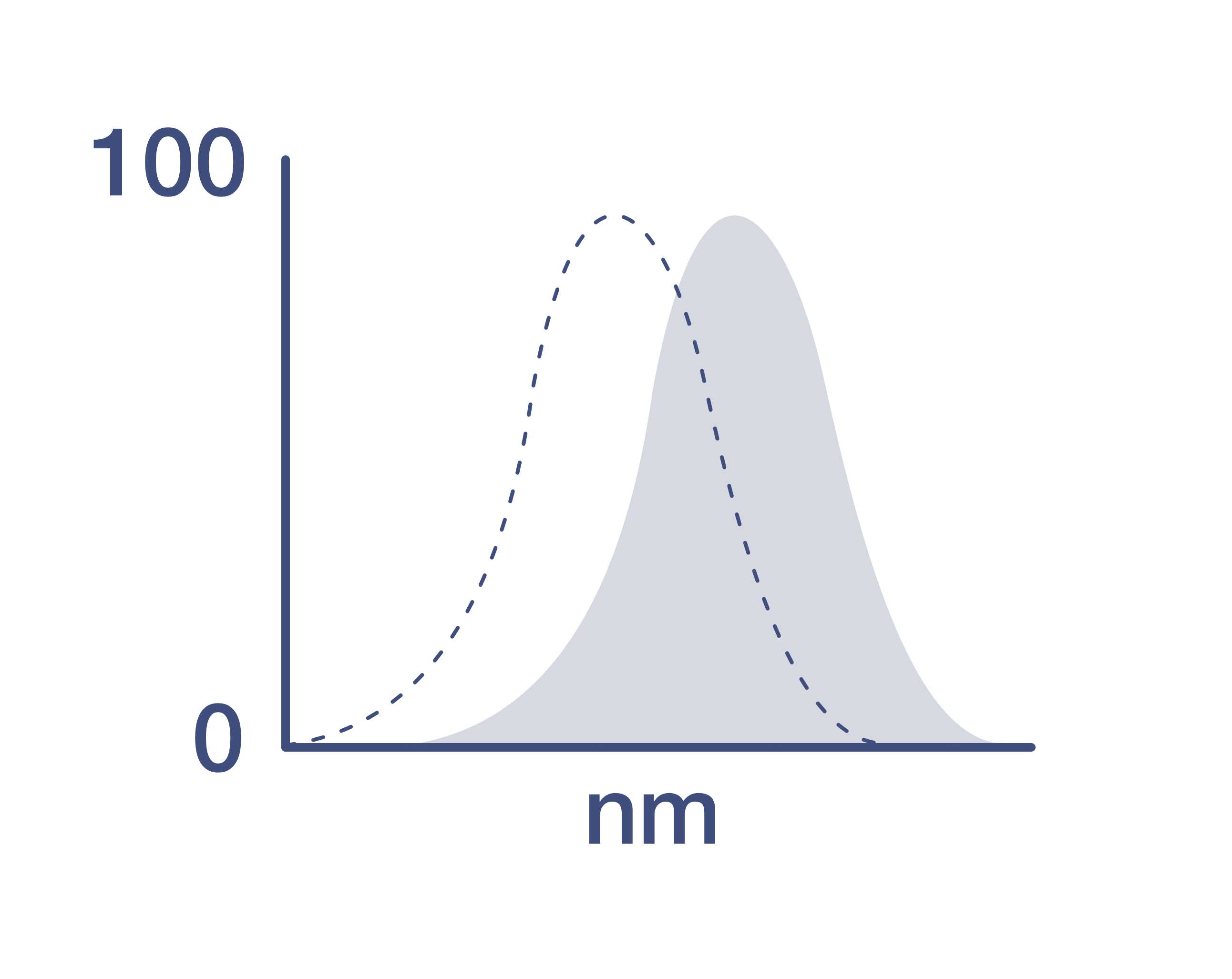
激发/发射光谱
482/708 nm
查看光谱
形式
Liquid
浓度
5 µL/Test
纯化类型
Affinity chromatography
保存液
PBS, pH 7.2, with BSA
内含物
0.09% sodium azide
保存条件
4°C, store in dark, DO NOT FREEZE!
运输条件
Ambient (domestic); Wet ice (international)
RRID
产品详细信息
Description: This 4H9 monoclonal antibody reacts with human CD7 (also known as Leu-9 or gp40), a 40-kDa transmembrane glycoprotein belonging to the immunoglobulin superfamily. This molecule is expressed on peripheral blood T lymphocytes and natural killer cells, as well as on lymphoid and myeloid lineage precursors during adult hematopoiesis. Capable of providing a costimulatory signal and associating with CD3 and CD45, CD7 is involved in T cell activation.
CD7 has also been detected on numerous T cell-derived leukemias and lymphomias. In fact, because CD7 expression is greater on CD4+ T cells in classical Hodgkin's lymphoma than in benign reactive lymph adenopathy, this molecule may be used as a diagnostic for Hodgkin's lymphoma.
Blocking assays demonstrate that this clone recognizes the same epitope as clone 124-1D1.
Applications Reported: This 4H9 antibody has been reported for use in flow cytometric analysis.
Applications Tested: This 4H9 antibody has been pre-titrated and tested by flow cytometric analysis of normal human peripheral blood cells. This can be used at 5 µL (0.25 µg) per test. A test is defined as the amount (µg) of antibody that will stain a cell sample in a final volume of 100 µL. Cell number should be determined empirically but can range from 10^5 to 10^8 cells/test.
PerCP-eFluor® 710 emits at 710 nm and is excited with the blue laser (488 nm); it can be used in place of PerCP-Cyanine5.5. We recommend using a 710/50 bandpass filter, however, the 695/40 bandpass filter is an acceptable alternative. Please make sure that your instrument is capable of detecting this fluorochrome.
Fixation: Samples can be stored in IC Fixation Buffer (Product # 00-8222) (100 µL cell sample + 100 µL IC Fixation Buffer) or 1-step Fix/Lyse Solution (Product # 00-5333) for up to 3 days in the dark at 4°C with minimal impact on brightness and FRET efficiency/compensation. Some generalizations regarding fluorophore performance after fixation can be made, but clone specific performance should be determined empirically.
Excitation: 488 nm; Emission: 710 nm; Laser: Blue Laser.
Filtration: 0.2 µm post-manufacturing filtered.
靶标信息
CD7, also known as gp40 or Leu9, is a 40 kDa receptor and member of the immunoglobulin gene superfamily. It features an N-terminal region (amino acids 1-107) that is highly homologous to Ig kappa light chains, while its carboxyl-terminal region is proline-rich, forming a stalk from which the Ig domain projects. CD7 is prominently expressed on the majority of immature and mature T lymphocytes, as well as T cell leukemias. It is also found on natural killer cells, a small subpopulation of normal B cells, and malignant B cells. CD7 plays a crucial role in modulating immune cell activity. Cross-linking of surface CD7 enhances T cell and NK cell functions, as evidenced by increased calcium flux, expression of adhesion molecules, cytokine secretion, and proliferation. CD7 directly associates with phosphoinositol 3-kinase, and its ligation induces the production of D-3 phosphoinositides and tyrosine phosphorylation. The expression of CD7 is an important marker in leukemia diagnostics, highlighting its significance in both normal immune function and disease states.
仅用于科研。不用于诊断过程。未经明确授权不得转售。
How to use the Panel Builder
Watch the video to learn how to use the Invitrogen Flow Cytometry Panel Builder to build your next flow cytometry panel in 5 easy steps.
生物信息学
蛋白别名: CD7; CD7 antigen (p41); GP40; p41 protein; T-cell antigen CD7; T-cell leukemia antigen; T-cell surface antigen Leu-9; TP41; unnamed protein product
基因别名: CD7; GP40; LEU-9; Tp40; TP41
Entrez Gene ID: (Human) 924
Disclaimer

Clicking the images or links will redirect you to a website hosted by BenchSci that provides third-party scientific content. Neither the content nor the BenchSci technology and processes for selection have been evaluated by us; we are providing them as-is and without warranty of any kind, including for use or application of the Thermo Fisher Scientific products presented.

