Search
Additional Information:
{{banner.description}}
")
图: 1 / 5
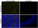
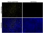
CD8a Antibody (740029TP555) in ICC/IF
Immunofluorescent analysis of CD8a was performed using CD8a transfected HEK-293E cells. The cells were fixed with 4% paraformaldehyde for 10 minutes, permeabilized with 0.1% Triton X-100 for 15 minutes and blocked with 2% BSA for 1 hour at room temperature. The cells were stained with CD8a Recombinant Rat Monoclonal Antibody (4SM16), Alexa Fluor™ Plus 555 (Product # 740029TP555, 1:200). Panel a) shows representative images of cells that were stained for detection and localization of CD8a. Panel c) is a composite image of the panel (a) and ... View More


Please note: We are reviewing Western blot images included in the antibody testing data in our catalog, including those provided by third parties. Unless expressly labeled or annotated as “raw-unedited”, Western blot images included in the antibody testing data in our catalog may have been edited, optimized or otherwise adjusted for presentation.
 in ICC/IF")
 in IHC (P)")
 in IHC (P)")
 in Flow")
")
产品信息
740029TP555
产品规格
种属反应
Human,
Mouse
宿主/亚型
Rat
/ IgG2a, kappa
Expression System
Expi293
分类
Recombinant Monoclonal
类型
Antibody
克隆号
4SM16
抗原
Fragment of extracellular domain
偶联物
Alexa Fluor™ Plus 555
Alexa Fluor™ Plus 555
Alexa Fluor™ Plus 555
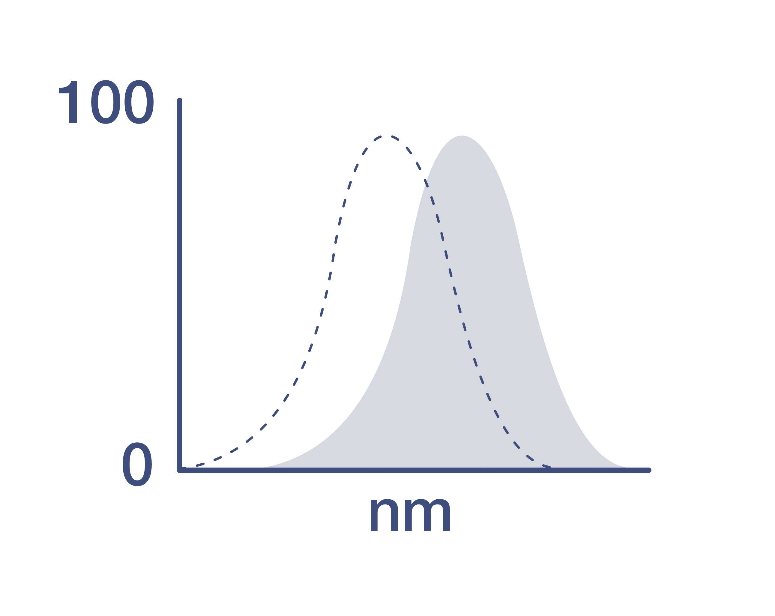
激发/发射光谱
558/572 nm
查看光谱
形式
Liquid
浓度
1.0 mg/mL
规格
50 µg
纯化类型
Affinity chromatography
保存液
proprietary buffer, pH 6.8
内含物
0.008% Bromonitrodioxane, 0.008% Methylisothiazolone
保存条件
4°C, store in dark, DO NOT FREEZE!
运输条件
Wet ice
RRID
产品详细信息
Alexa Fluor™ Plus recombinant rat antibodies are conjugated using new, proprietary dye chemistry so you can generate stunning data. Alexa Fluor™ Plus antibodies represent an advancement in fluorescent conjugate technology. Alexa Fluor™ Plus antibodies provide brighter signal compared to leading Alexa Fluor™ antibodies, providing you with better signal-to-noise for your critical experiments. These antibodies show better specificity and lot-to-lot consistency as these are recombinant antibodies, generated by cloning specific genes for the desired antibodies into an expression vector and expressed in vitro.
Using conjugate solutions: Centrifuge the protein conjugate solution briefly in a microcentrifuge before use; add only the supernatant to the experiment. This step will help eliminate any protein aggregates that may have formed during storage, thereby reducing nonspecific background staining.
Applications Tested: This 4SM16 antibody has been tested by immunohistochemistry, immunocytochemistry and flow cytometric analysis. This may be used for immunohistochemistry at 20 µg/mL, for immunocytochemistry at 5 µg/mL at and for flow cytometry at less than or equal to 0.5 µg per test. A test is defined as the amount (µg) of antibody that will stain a cell sample in a final volume of 100 µL. Cell number should be determined empirically but can range from 10^5 to 10^8 cells/test. It is recommended that the antibody be carefully titrated for optimal performance in the assay of interest.
Excitation: 553 nm; Emission: 568 nm; Laser: Yellow Laser
Filtration: 0.2 µm post-manufacturing filtered.
靶标信息
CD8, also known as cluster of differentiation 8, is a type I transmembrane glycoprotein of the immunoglobulin family that plays a crucial role in T cell differentiation, activation, and signal transduction. It is expressed as either a heterodimer (CD8 alpha beta) or a homodimer (CD8 alpha alpha). The CD8 alpha beta form is predominantly found on the majority of thymocytes and a subpopulation of mature alpha beta TCR T cells, while the CD8 alpha alpha form is expressed on gamma delta TCR T cells, a subset of intestinal intraepithelial lymphocytes (IELs), and dendritic cells. CD8 functions as a co-receptor for major histocompatibility complex class I (MHC-I) molecules, working alongside the T cell receptor (TCR). The CD8 alpha chain is essential for binding to MHC-I. CD8 is also expressed on a subset of T cells, NK cells, monocytes, and dendritic cells as disulfide-linked homodimers of CD8 alpha. Upon ligation of MHC-I/peptide complexes presented by antigen-presenting cells (APCs), CD8 recruits lymphocyte-specific protein tyrosine kinase (Lck), leading to lymphokine production, increased motility, and activation of cytotoxic T lymphocytes (CTLs). Activated CTLs are vital for clearing pathogens and tumor cells. The differentiation of naive CD8+ T cells into CTLs is strongly enhanced by cytokines such as IL-2, IL-12, and TGF-beta1. Through its interactions with MHC-I and association with protein tyrosine kinase p56lck, CD8 plays a significant role in T cell development and the activation of mature T cells.
仅用于科研。不用于诊断过程。未经明确授权不得转售。
篇参考文献 (0)
您是否在文献中引用过该产品?请点击下方按钮邮件告知我们。
生物信息学
蛋白别名: CD8a; CD8alpha; CD8b; CD8beta; fCD8; Leu-2; leu-2a; Lymphocyte antigen 3; Lyt-2.1 lymphocyte differentiation antigen (AA at 100); T-cell membrane glycoprotein Ly-3; T-cell surface glycoprotein CD8 alpha chain; T-cell surface glycoprotein CD8 beta chain; T-cell surface glycoprotein Lyt-2; T-cell surface glycoprotein Lyt-3; T-lymphocyte differentiation antigen T8/Leu-2
基因别名: BB154331; CD8A; CD8B; CD8B1; Ly-2; Ly-3; Ly-35; Ly-B; Ly-C; Lyt-2; Lyt-3; Lyt2; Lyt3; MAL
UniProt ID: (Mouse) P01731, (Mouse) P10300
Entrez Gene ID: (Mouse) 12525, (Mouse) 12526
Disclaimer

Clicking the images or links will redirect you to a website hosted by BenchSci that provides third-party scientific content. Neither the content nor the BenchSci technology and processes for selection have been evaluated by us; we are providing them as-is and without warranty of any kind, including for use or application of the Thermo Fisher Scientific products presented.

