Search
Invitrogen
CD18 (LFA-1 beta) Monoclonal Antibody (6.7), NovaFluor™ Blue 660-120S, eBioscience™
{{$productOrderCtrl.translations['antibody.pdp.commerceCard.promotion.promotions']}}
{{$productOrderCtrl.translations['antibody.pdp.commerceCard.promotion.viewpromo']}}
{{$productOrderCtrl.translations['antibody.pdp.commerceCard.promotion.promocode']}}: {{promo.promoCode}} {{promo.promoTitle}} {{promo.promoDescription}}. {{$productOrderCtrl.translations['antibody.pdp.commerceCard.promotion.learnmore']}}
Additional Information:
{{banner.description}}
 Antibody in Flow Cytometry (Flow)")
图: 1 / 2
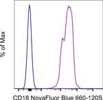
CD18 (LFA-1 beta) Antibody (H083T03B08-A) in Flow
Normal human peripheral blood cells were either left unstained (blue histogram) or stained with CD18 (LFA-1 beta) Monoclonal Antibody, NovaFluor Blue 660-120S (purple histogram). Total viable cells in the lymphocyte gate were used for analysis, as determined by LIVE/DEAD Blue (Product # L34962). Data was acquired on a 5-laser Cytek Aurora and unmixed with autofluorescence extraction.
Please note: We are reviewing Western blot images included in the antibody testing data in our catalog, including those provided by third parties. Unless expressly labeled or annotated as “raw-unedited”, Western blot images included in the antibody testing data in our catalog may have been edited, optimized or otherwise adjusted for presentation.
 Antibody (H083T03B08-A) in Flow")
 Antibody (H083T03B08-A) in Flow")
产品信息
H083T03B08-A
产品规格
种属反应
Human
宿主/亚型
Mouse
/ IgG1, kappa
分类
Monoclonal
类型
Antibody
克隆号
6.7
偶联物
NovaFluor™ Blue 660-120S
NovaFluor™ Blue 660-120S
NovaFluor™ Blue 660-120S
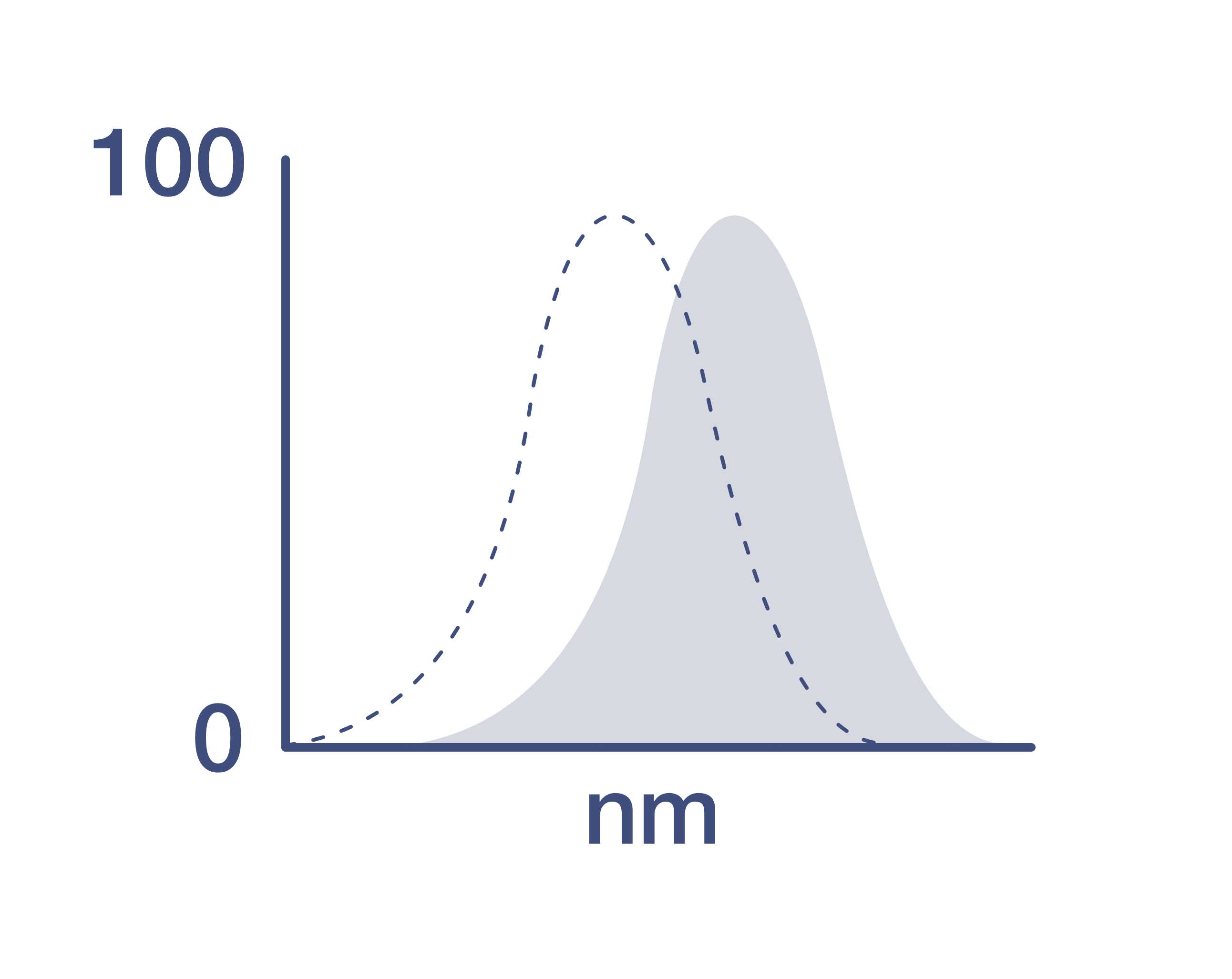
激发/发射光谱
492/665 nm
查看光谱
形式
Liquid
浓度
0.8 µg/Test
纯化类型
Affinity chromatography
保存液
PBS, pH 7.2, with BSA
内含物
0.09% sodium azide
保存条件
4°C, store in dark, DO NOT FREEZE!
RRID
产品详细信息
Description
The 6.7 monoclonal antibody reacts with human and non-human primate (rhesus or cynomologus) CD18, which is also known as beta2 integrin. CD18 is expressed broadly by human lymphocytes and monocytes. Peripheral blood granulocytes also express CD18, but at lower levels. The 6.7 monoclonal antibody has been reported to induce the proliferation of resting T cells cultured with monocytes.
This product contains 1 vial of NovaFluor conjugate and 1 vial of CellBlox Plus Blocking Buffer.
Applications Tested
This 6.7 antibody has been pre-titrated and tested by flow cytometric analysis of normal human blood cells. This can be used at 5 µL (0.8 µg) per test. A test is defined as the amount (µg) of antibody that will stain a cell sample in a final volume of 100 µL. Cell number should be determined empirically but can range from 10^5 to 10^8 cells/test.
Master mixes
• Master mixes of NFs should be made at 2-8 °C and may be made up to 4 hours ahead of time.
• We do not recommend storing master mixes containing NovaFluor conjugates overnight or longer.
Whole Blood compatibility
• When utilizing whole blood (as opposed to density-gradient-purified PBMC), we recommend lysing red blood cells in bulk prior to staining with NovaFluor conjugates.
• See the Bulk Lysis of Human Whole Blood protocol here.
• Staining of whole blood with NovaFluor conjugates followed by lysis of red blood cells may result in higher-than-expected background staining.
Viability dye compatibility
• NovaFluor dyes are not compatible with DNA intercalating viability dyes.
• Do not use viability dyes such as propidium iodide, 7-actinomycin D (7-AAD) and DAPI. Invitrogen LIVE/DEAD Fixable Dead Cell stains are recommended for use with NovaFluor dyes.
CellBlox Plus Blocking Buffer
• This NovaFluor conjugate comes with CellBlox Plus Blocking Buffer (for 25 µg or 25 test products, Cat. No. C001T02F01; for 100 µg or 100 test, Cat. No. C001T03F01), essential for optimal staining.
• Use CellBlox Plus Blocking Buffer in all experiments with NovaFluor conjugates.
• Add 5 µL per sample to antibody cocktails/master mixes (regardless of how many Novafluor-conjugated antibodies are present) before combining with cells.
• CellBlox Plus Blocking Buffer is compatible with either Super Bright Complete Blocking Buffer or Brilliant Stain Buffer and can be used in antibody cocktails/master mixes with those reagents.
• For single-color controls, use 5 µL of CellBlox Plus Blocking Buffer per 100 µL of cell sample (10^3 to 10^8 cells).
NovaFluor conjugates are based on Phiton technology utilizing novel fluorophore-containing nucleic acid dye structures that allow for engineered fluorescent signatures with consideration for spillover and spread impacts. Learn more
Excitation: 508 nm; Emission: 664 nm; Laser: 488 nm (Blue) Laser
靶标信息
CD18, also known as the integrin beta 2 subunit, is a 90-95 kDa type I transmembrane protein expressed on all leukocytes. It heterodimerizes with the alpha chains of CD11a, CD11b, CD11c, and CD11d to form various leukocyte (beta2) integrins, which are crucial for cellular adhesion and signaling. The CD18/CD11a complex, known as lymphocyte function-associated antigen-1 (LFA-1), plays a significant role in cellular adhesion and inflammatory reactions. CD18 also forms heterodimers with CD11b (Mac-1, CR3), CD11c, and CD11d, contributing to the processes of cell adhesion and cell-surface mediated signaling. These integrins are essential for proper leukocyte migration and mediating intercellular contacts. The absence of CD18 leads to leukocyte adhesion deficiency type I (LAD1), a condition characterized by impaired leukocyte migration. Severe reduction in CD18 expression can result in the development of a psoriasiform skin disease. CD18 is also a target of Mannheimia (Pasteurella) haemolytica leukotoxin, which can mediate leukotoxin-induced cytolysis. Defects in the CD18 gene are the cause of LAD1, and two transcript variants encoding the CD18 protein have been identified. The integrin-mediated responses are often a composite of the functions of its individual subunits, highlighting the importance of CD18 in immune function and disease.
仅用于科研。不用于诊断过程。未经明确授权不得转售。
How to use the Panel Builder
Watch the video to learn how to use the Invitrogen Flow Cytometry Panel Builder to build your next flow cytometry panel in 5 easy steps.
篇参考文献 (0)
您是否在文献中引用过该产品?请点击下方按钮邮件告知我们。
生物信息学
蛋白别名: cell surface adhesion glycoprotein (LFA-1/CR3/P150,959 beta subunit precursor); complement component 3 receptor 3 and 4 subunit; complement receptor C3 beta-subunit; integrin beta chain, beta 2; integrin, beta 2 (complement component 3 receptor 3 and 4 subunit); leukocyte adhesion protein beta-subunit precursor; leukocyte cell adhesion molecule CD18; leukocyte-associated antigens CD18/11A, CD18/11B, CD18/11C; putative; unnamed protein product
基因别名: CD18; LAD; LCAMB; LFA-1; MAC-1; MF17; MFI7
UniProt ID: (Human) P05107
Entrez Gene ID: (Human) 3689
microglial cell activation
leukocyte migration involved in inflammatory response
receptor-mediated endocytosis
phagocytosis
phagocytosis, engulfment
apoptotic process
inflammatory response
cell adhesion
neuron cell-cell adhesion
leukocyte cell-cell adhesion
cell-matrix adhesion
integrin-mediated signaling pathway
I-kappaB kinase/NF-kappaB signaling
cell-cell signaling
regulation of cell shape
neutrophil chemotaxis
receptor internalization
positive regulation of superoxide anion generation
cell adhesion mediated by integrin
cell-cell adhesion mediated by integrin
heterotypic cell-cell adhesion
endodermal cell differentiation
receptor clustering
positive regulation of neutrophil degranulation
endothelial cell migration
cellular extravasation
positive regulation of nitric oxide biosynthetic process
positive regulation of angiogenesis
negative regulation of dopamine metabolic process
anatomical structure formation involved in morphogenesis
regulation of peptidyl-tyrosine phosphorylation
leukocyte adhesion to vascular endothelial cell
cellular response to low-density lipoprotein particle stimulus
positive regulation of protein targeting to membrane
beta-amyloid clearance
cell-cell adhesion
positive regulation of leukocyte adhesion to vascular endothelial cell
neutrophil migration
Disclaimer

Clicking the images or links will redirect you to a website hosted by BenchSci that provides third-party scientific content. Neither the content nor the BenchSci technology and processes for selection have been evaluated by us; we are providing them as-is and without warranty of any kind, including for use or application of the Thermo Fisher Scientific products presented.

