Search
Invitrogen
P-cadherin Monoclonal Antibody (PCD-1)
{{$productOrderCtrl.translations['antibody.pdp.commerceCard.promotion.promotions']}}
{{$productOrderCtrl.translations['antibody.pdp.commerceCard.promotion.viewpromo']}}
{{$productOrderCtrl.translations['antibody.pdp.commerceCard.promotion.promocode']}}: {{promo.promoCode}} {{promo.promoTitle}} {{promo.promoDescription}}. {{$productOrderCtrl.translations['antibody.pdp.commerceCard.promotion.learnmore']}}
Additional Information:
{{banner.description}}
")
图: 1 / 11
P-cadherin Antibody (13-2000Z) in IHC
Current issues in molecular biology 2023 -
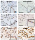
Representative microphotographs of immunohistochemical staining using antibodies against E-cadherin (E-cad) (DAKO, M3612, NCH-38) ( A ), E-cadherin (ProteinTech, 20874-1-AP) ( B ), N-cadherin (N-cad) ( C ), P-cadherin (P-cad) (Santa Cruz, sc-74545) ( D ), beta-catenin ( E ), and vimentin ( F ). Insets of ( A - F ) show enlarged images of microphotographs. Images from cases 1 ( F ), 3 ( C ), 5 ( E ), 6 ( A )... View More


 in IHC")
 in ICC/IF")
 in ICC/IF")
 in IHC (P)")
 in WB, IHC (P)")
 in IHC (P)")
 in IHC (F)")
 in IHC")
 in IHC")
 in WB")
 in WB")
产品信息
13-2000Z
应用
建议稀释比
已发表文章
产品规格
种属反应
Mouse
已发表种属
Bovine,
Fish,
Hamster,
Human,
Mouse,
Rat,
Sheep,
Tag
宿主/亚型
Rat
/ IgG2a
分类
Monoclonal
类型
Antibody
克隆号
PCD-1
抗原
Mouse endoderm cell line PSA5-E.
偶联物
形式
Lyophilized
浓度
2.0 mg/mL
规格
100 µg
纯化类型
Ion-exchange chromatography
保存液
PBS, pH 7.4, with 1% BSA
内含物
no preservative
保存条件
4°C
运输条件
Ambient (domestic); Wet ice (international)
RRID
产品详细信息
Source: Monoclonal antibody was obtained by fusing the P3-X63-Ag8-U1 mouse myeloma cell-line with spleen cells of Donryu rat after immunization with mouse endoderm cell line PSA5-E.
Specificity: This antibody specifically reacts with mouse P-cadherin. This antibody inhibits P-cadherin-dependent cell-cell contact.
Cross reactivity: This antibody does not react with human or chicken P-cadherin.
Storage: 4°C This product does not contain preservative. The stock solution (2.0 mg/ml) should be stored in aliquots at -20°C for 1 year, or should be stored at 4°C for 6 months after adding 0.1% sodium azide. Avoid repeated freeze-thaw cycles.
Reconstitution: Dissolve the lyophilized antibody in 50 µl of distilled water (final concentration: 2.0 mg/ml). This solution can be used as a stock solution. If dilution is necessary for your application, dilute the stock solution with the following Dilution solution just prior to use. When the entire amount of antibody is to be used over a short time period, it may be dissolved directly in 500 µl or more of the Dilution solution. Be sure to store the antibody at a minimum concentration of 2.0 mg/ml. A lower antibody concentration may result in decreased stability. Reconstituted antibody solution should contain 0.1% sodium azide as a preservative when stored at 4°C.
Ca2+ should be contained in the buffers to stabilize cadherin antigen.
靶标信息
P-cadherin, also known as Cadherin-3, is a Type 1 cadherin protein that belongs to the cadherin superfamily. Type 1 cadherins are single-pass transmembrane proteins that have 5 extracellular cadherin repeats and an intracellular domain that binds p120-catenin and beta-catenin. Cadherins are calcium-dependent cell-cell adhesion glycoproteins responsible for a range of processes including development, wound healing, cell-cell signaling, cell growth and differentiation. P-cadherin is expressed in human fetal structures, and in adult tissues such as the basal layer of the epidermis, breast, prostate, ovary, cervix, hair follicle, and corneal endothelium. P-cadherin expression has also been reported on embryonic stem cells, and stem cells of the normal mammary gland, and hair follicle. Overexpression of P-cadherin has been associated with poor prognosis in breast, prostate, ovary, colon, and stomach carcinomas. Mutations in CDH3 are associated with congential hypotrichosis with juvenile macular dystrophy.
仅用于科研。不用于诊断过程。未经明确授权不得转售。
生物信息学
蛋白别名: CADH3; Cadherin-3; P-cadherin; Placental cadherin; Placental-cadherin; RPE-specific cadherin
基因别名: AI385538; Cadp; Cdh3; Cdhp; P-cadherin; Pcad
UniProt ID: (Mouse) P10287
Entrez Gene ID: (Mouse) 12560
retina homeostasis
cell adhesion
homophilic cell adhesion via plasma membrane adhesion molecules
positive regulation of gene expression
positive regulation of keratinocyte proliferation
single organismal cell-cell adhesion
hair cycle process
keratinization
positive regulation of monophenol monooxygenase activity
negative regulation of transforming growth factor beta2 production
positive regulation of insulin-like growth factor receptor signaling pathway
positive regulation of melanin biosynthetic process
negative regulation of catagen
canonical Wnt signaling pathway
regulation of hair cycle by canonical Wnt signaling pathway
positive regulation of melanosome transport
Disclaimer

Clicking the images or links will redirect you to a website hosted by BenchSci that provides third-party scientific content. Neither the content nor the BenchSci technology and processes for selection have been evaluated by us; we are providing them as-is and without warranty of any kind, including for use or application of the Thermo Fisher Scientific products presented.

