Search
Invitrogen
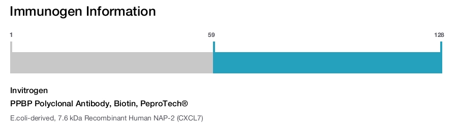
PPBP Polyclonal Antibody, Biotin, PeproTech®
{{$productOrderCtrl.translations['antibody.pdp.commerceCard.promotion.promotions']}}
{{$productOrderCtrl.translations['antibody.pdp.commerceCard.promotion.viewpromo']}}
{{$productOrderCtrl.translations['antibody.pdp.commerceCard.promotion.promocode']}}: {{promo.promoCode}} {{promo.promoTitle}} {{promo.promoDescription}}. {{$productOrderCtrl.translations['antibody.pdp.commerceCard.promotion.learnmore']}}
Additional Information:
{{banner.description}}
")
图: 1 / 3
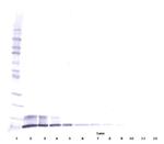
PPBP Antibody (500-P03GBT-25UG) in WB
Western Blot: To detect Human NAP-2 by Western Blot analysis PPBP Polyclonal Antibody, Biotin (Product # 500-P03GBT-500UG) can be used at a concentration of 0.1-0.2 µg/mL. When used in conjunction with compatible development reagents the detection limit for Recombinant Human NAP-2 is 1.5-3.0 ng/lane, under either reducing or non-reducing conditions.
Please note: We are reviewing Western blot images included in the antibody testing data in our catalog, including those provided by third parties. Unless expressly labeled or annotated as “raw-unedited”, Western blot images included in the antibody testing data in our catalog may have been edited, optimized or otherwise adjusted for presentation.
 in WB")
 in WB")
 in ELISA")
产品信息
500-P03GBT-25UG
产品规格
种属反应
Human
宿主/亚型
Goat
分类
Polyclonal
类型
Antibody
抗原
E.coli-derived, 7.6 kDa Recombinant Human NAP-2 (CXCL7)
偶联物
Biotin
Biotin
Biotin
形式
Lyophilized
浓度
0.1-1.0 mg/mL
规格
25 µg
纯化类型
Antigen affinity chromatography
保存液
PBS
内含物
no preservative
保存条件
-20°C
运输条件
Ambient
RRID
产品详细信息
AA Sequence of recombinant protein: AELRCMCIKT TSGIHPKNIQ SLEVIGKGTH CNQVEVIATL KDGRKICLDP DAPRIKKIVQ KKLAGDESAD.
Preparation: Produced from sera of goats immunized with highly pure Recombinant Human NAP-2 (CXCL7). Anti-Human NAP-2 (CXCL7)-specific antibody was purified by affinity chromatography and then biotinylated.
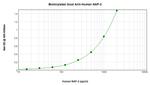
Sandwich ELISA: To detect Human NAP-2 by sandwich ELISA (using 100 µL/well) a concentration of 0.25-1.0 µg/mL of this antibody is required. This biotinylated polyclonal antibody, in conjunction with PeproTech Polyclonal Anti-Human NAP-2 (CXCL7) (500-P03) as a capture antibody, allows the detection of at least 2000-4000 pg/mL of Recombinant Human NAP-2.
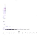
Western Blot: To detect Human NAP-2 by Western Blot analysis this antibody can be used at a concentration of 0.1-0.2 µg/mL. When used in conjunction with compatible development reagents the detection limit for Recombinant Human NAP-2 is 1.5-3.0 ng/lane, under either reducing or non-reducing conditions.
500-P03GBT-1MG will be provided as 2 x 500 µg
靶标信息
Members of the a-chemokine subfamily of inducible, secreted, pro-inflammatory cytokines contain a similar motif, in which the first two cysteine residues are separated by a single residue (Cys-X-Cys), and are also chemotactic for neutrophils. The platelet basic protein (PBP), a member of the a-chemokine family, resides in the a-granules of platelets and is released upon their activation. Proteolytic cleavage of the amino terminus of PBP leads to the generation of several peptides, which include mature PBP, connective tissue-activating peptide III (CTAP III, also designated low affinity platelet factor IV (LA-PF4)), b-thromboglobulin (b-TG), and neutrophil-activating peptide 2 (NAP-2). PBP and its N-truncated derivatives mediate inflammation and wound healing. Specifically, NAP-2 activates chemotaxis and degranulation in neutrophils during inflammation. The gene encoding human PBP maps to chromosome 4q12-q13.
仅用于科研。不用于诊断过程。未经明确授权不得转售。
篇参考文献 (0)
您是否在文献中引用过该产品?请点击下方按钮邮件告知我们。
生物信息学
蛋白别名: C-X-C motif chemokine 7; LDGF; Leukocyte-derived growth factor; Macrophage-derived growth factor; MDGF; PBP; Platelet basic protein; Small-inducible cytokine B7
基因别名: CTAP3; CXCL7; PPBP; SCYB7; TGB1; THBGB1
Entrez Gene ID: (Human) 5473
Disclaimer

Clicking the images or links will redirect you to a website hosted by BenchSci that provides third-party scientific content. Neither the content nor the BenchSci technology and processes for selection have been evaluated by us; we are providing them as-is and without warranty of any kind, including for use or application of the Thermo Fisher Scientific products presented.

