Search
Invitrogen
PRAME/CD3 (Brenetafusp Biosimilar) Recombinant Human Monoclonal Antibody
{{$productOrderCtrl.translations['antibody.pdp.commerceCard.promotion.promotions']}}
{{$productOrderCtrl.translations['antibody.pdp.commerceCard.promotion.viewpromo']}}
{{$productOrderCtrl.translations['antibody.pdp.commerceCard.promotion.promocode']}}: {{promo.promoCode}} {{promo.promoTitle}} {{promo.promoDescription}}. {{$productOrderCtrl.translations['antibody.pdp.commerceCard.promotion.learnmore']}}
Additional Information:
{{banner.description}}
 Antibody in SDS-PAGE (SDS-PAGE)")
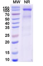
 Antibody (MA562571) in SDS-PAGE")
产品信息
MA562571
应用
建议稀释比
已发表文章
产品规格
种属反应
Human
宿主/亚型
Human
/ Ig Mix
Expression System
CHO cells
分类
Recombinant Monoclonal
类型
Antibody
偶联物
Unconjugated
Unconjugated
Unconjugated
形式
Liquid
浓度
1 mg/mL
规格
100 µg
纯化类型
Metal-chelate chromatography
保存液
PBS, pH 7.4
内含物
no preservative
保存条件
Store at 4°C short term. For long term storage, store at -20°C, avoiding freeze/thaw cycles.
运输条件
Ambient (domestic); Wet ice (international)
RRID
靶标信息
The PRAME gene (Preferentially Expressed Antigen in Melanoma) is located on chromosome 22q11.22. It encodes a protein involved in the immune response against cancer cells, particularly in melanoma. The PRAME protein consists of multiple leucine-rich repeat (LRR) motifs, which play a role in protein-protein interactions. Functionally, PRAME acts as a repressor of retinoic acid (RA) signaling by binding to RA receptor targets and inhibiting their transcriptional activation. This inhibition of RA signaling contributes to the proliferation and survival of cancer cells, making PRAME a significant tumor-associated antigen and a potential target for cancer immunotherapy. Elevated expression of PRAME has been observed in various cancers, including melanoma, lung cancer, and renal cell carcinoma.
The CD3 complex, composed of gamma, delta, epsilon, and zeta subunits, is essential for the assembly, trafficking, and surface expression of the T cell receptor (TCR) complex. These subunits are structurally related members of the immunoglobulin superfamily and are encoded by closely linked genes on human chromosome 11. CD3 is expressed by thymocytes in a developmentally regulated manner and by all mature T cells, but not on B or NK cells. The CD3 subunits play a crucial role in transducing antigen-recognition signals into the cytoplasm of T cells. The cytoplasmic tails of CD3 subunits contain a double tyrosine-based motif that associates with cytoplasmic signal transduction molecules, mediating T cell activation through the TCR. Crosslinking of the TCR initiates intracellular biochemical pathways that result in cellular activation, proliferation, and potentially growth arrest and cell survival. CD3 is present on 68-82% of normal peripheral blood lymphocytes, 65-85% of thymocytes, and Purkinje cells in the cerebellum. Decreased percentages of T lymphocytes may be observed in some autoimmune diseases. Defects in the CD3 gene are associated with CD3 immunodeficiency, highlighting its importance in immune function and regulation.
仅用于科研。不用于诊断过程。未经明确授权不得转售。
篇参考文献 (0)
您是否在文献中引用过该产品?请点击下方按钮邮件告知我们。
Disclaimer

Clicking the images or links will redirect you to a website hosted by BenchSci that provides third-party scientific content. Neither the content nor the BenchSci technology and processes for selection have been evaluated by us; we are providing them as-is and without warranty of any kind, including for use or application of the Thermo Fisher Scientific products presented.

