Search
Invitrogen
Perforin Monoclonal Antibody (eBioOMAK-D), PE, eBioscience™
{{$productOrderCtrl.translations['antibody.pdp.commerceCard.promotion.promotions']}}
{{$productOrderCtrl.translations['antibody.pdp.commerceCard.promotion.viewpromo']}}
{{$productOrderCtrl.translations['antibody.pdp.commerceCard.promotion.promocode']}}: {{promo.promoCode}} {{promo.promoTitle}} {{promo.promoDescription}}. {{$productOrderCtrl.translations['antibody.pdp.commerceCard.promotion.learnmore']}}
Additional Information:
{{banner.description}}
")
图: 1 / 24
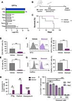
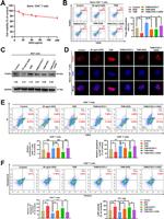
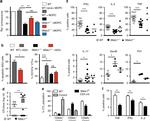
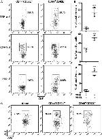
Perforin Antibody (12-9392-82) in Flow
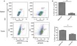
Intracellular staining of C57BL/6 splenocytes unstimulated (left) or stimulated with Mouse IL-2 Recombinant Protein (Product # 14-8021-64) (right) with Anti-Mouse CD49b (Integrin alpha 2) APC (Product # 17-5971-82) and 0.5 µg of Anti-Mouse Perforin PE using the Foxp3/Transcription Factor Staining Buffer Set (Product # 00-5523-00) and protocol. Quadrant lines demarcate isotype control staining.

Please note: We are reviewing Western blot images included in the antibody testing data in our catalog, including those provided by third parties. Unless expressly labeled or annotated as “raw-unedited”, Western blot images included in the antibody testing data in our catalog may have been edited, optimized or otherwise adjusted for presentation.
 in Flow")
 in Flow")
 in Flow")
 in Flow")
 in Flow")
 in Flow")
 in Flow")
 in Flow")
 in Flow")
 in Flow")
 in Flow")
 in Flow")
 in Flow")
 in Flow")
 in Flow")
 in Flow")
 in Flow")
 in Flow")
 in Flow")
 in Flow")
 in Flow")
 in Flow")
 in Flow")
")
产品信息
12-9392-82
应用
建议稀释比
已发表文章
产品规格
种属反应
Mouse
已发表种属
Human,
Mouse
宿主/亚型
Rat
/ IgG2a, kappa
分类
Monoclonal
类型
Antibody
克隆号
eBioOMAK-D
偶联物
PE
PE
PE
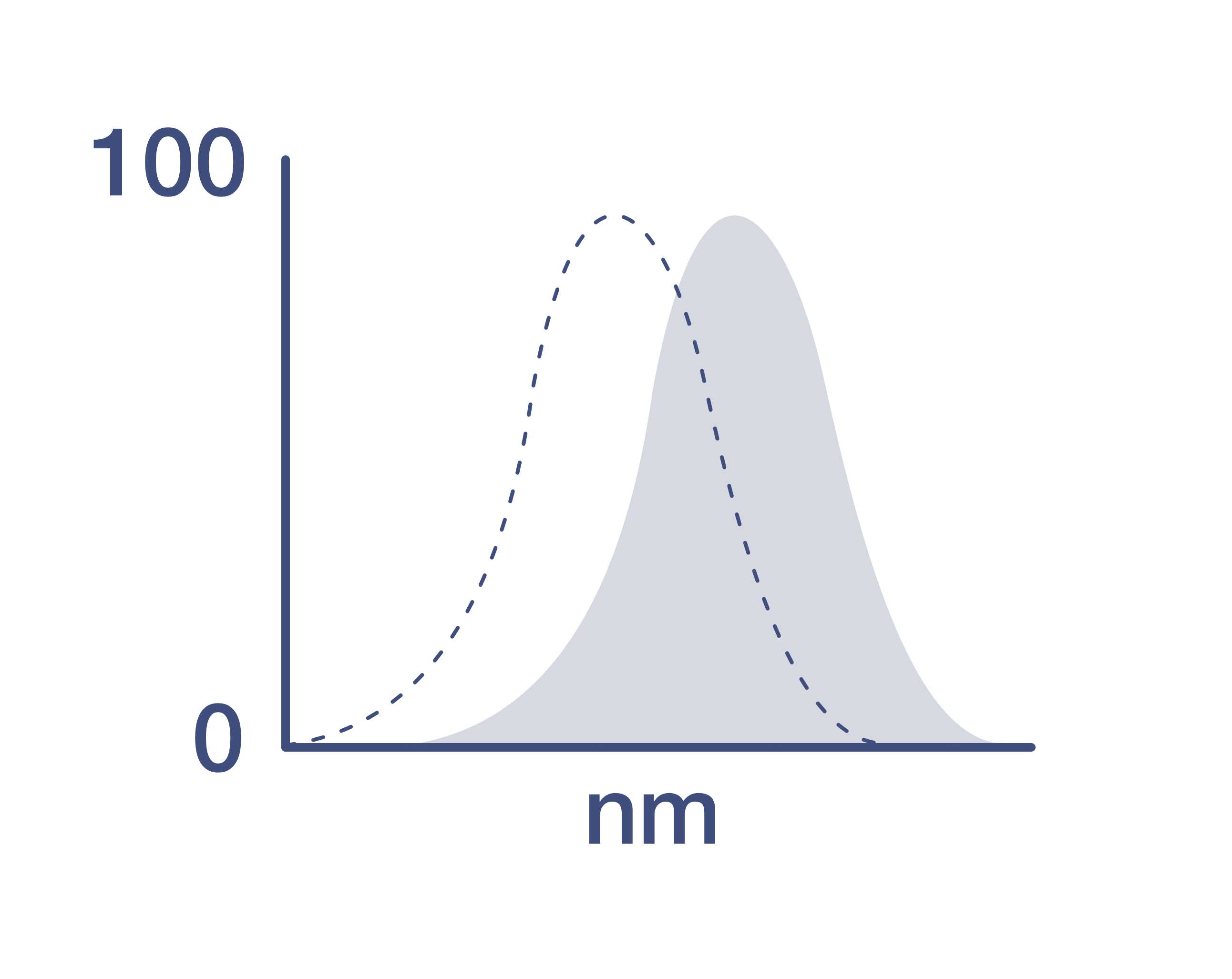
激发/发射光谱
565/576 nm
查看光谱
形式
Liquid
浓度
0.2 mg/mL
规格
100 µg
纯化类型
Affinity chromatography
保存液
PBS, pH 7.2
内含物
0.09% sodium azide
保存条件
4°C, store in dark, DO NOT FREEZE!
运输条件
Ambient (domestic); Wet ice (international)
RRID
产品详细信息
Description: The eBioOMAK-D antibody reacts with mouse perforin (pore-forming protein, pfp, Prf). Perforin is one of the cytolytic mediators present in the cytoplasmic granules of cytotoxic T lymphocytes (CTL) and natural killer cells (NK). Perforin is involved in the killing function by CTLs and NKs and has an important role in the immune response against tumors and virus infections.
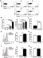
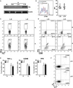
By immunoblotting, eBioOMAK-D recognizes a ~70kDa band in lysates of CTLL-2 mouse cytotoxic cell line and in lysates of IL-2 stimulated but not unstimulated mouse splenocytes. By multi-color intracellular flow cytometric analysis, eBioOMAK-D staining is increased upon stimulation (IL-2 or anti-CD3/28). Intracellular flow staining results showing upregulation of protein expression have been confirmed by immunoblotting. Furthermore, stimulated Perforin Knock-out (developed by Walsh) splenocytes do not stain with eBioOMAK-D nor is any protein detectable by western blotting with eBioOMAK-D as well as other anti-mouse perforin antibodies. Please note that the Kagi perforin knock-out mice may synthesize a truncated form of the protein which may be recognized by eBioOMAK-D.
In IL-2 stimulated mouse splenocytes, NK cells (as determined by CD49b staining) contain perforin while CD8 cells contain little to none and can vary with culture conditions. This has been confirmed by staining and western blotting the two populations using both OMAK-D and P1-8 antibodies. In contrast stimulation of splenocytes with anti-CD3/CD28 antibodies does result in an increase of perforin on both NK cells and CD8 cells.
eBioOMAK-D is also cross-reactive to human perforin and co-stains CD56 positive cells in PBMC.
Expression of perforin and Granzyme B do not always correlate (as discussed above in the CD8 population of IL-2 stimulated splenocytes). Granzyme B typically is expressed earlier and at higher levels. Expression of Granzyme B is dramatically increased (more than 10,00 fold based on mRNA estimates and significantly at the protein level based on western blotting and flow analysis) compared to a minimal increase (10-100 fold) in perforin mRNA and protein with IL-2 stimulation.
For intracellular staining and flow cytometric analysis with direct conjugates of anti-mouse perforin, it is highly recommended to use the Foxp3 buffer system (Product # 00-5523). Other buffers may yield varying results.
Applications Reported: This eBioOMAK-D antibody has been reported for use in intracellular staining followed by flow cytometric analysis.
Applications Tested: This eBioOMAK-D antibody has been tested by intracellular staining and flow cytometric analysis of stimulated mouse splenocytes using the Foxp3/Transcription Factor Staining Buffer Set (Product # 00-5523) and protocol. Please refer to BestProtocols®: Protocol B: One step protocol for intracellular (nuclear) proteins. This can be used at less than or equal to 1 µg per test. A test is defined as the amount (µg) of antibody that will stain a cell sample in a final volume of 100 µL. Cell number should be determined empirically but can range from 10^5 to 10^8 cells/test. It is recommended that the antibody be carefully titrated for optimal performance in the assay of interest.
Excitation: 488-561 nm; Emission: 578 nm; Laser: Blue Laser, Green Laser, Yellow-Green Laser.
Filtration: 0.2 µm post-manufacturing filtered.
靶标信息
Perforin is one of the major cytolytic proteins of cytolytic granules. Perforin is a cytolytic mediator and is stored in and released by cytoplasmic granules. Moreover, perforin is involved in immune defense against tumors and virus infections as mediated by cytotoxic lymphocytes. Perforin is a 555 amino acid protein with a 21 amino acid signal peptide, and has a molecular weight of 70 to 75 kD. Perforin is a pore forming protein with a mechanism of transmembrane channel formation similar to C9, and homology between perforin and C9 have been demonstrated. Studies show that perforin is expressed only in killer cell lines and not in helper T lymphocytes or other tumor cells tested. Perforin is known to be a key effector molecule for T-cell- and natural killer-cell-mediated cytolysis. Defects in the perforin gene cause familial hemophagocytic lymphohistiocytosis type 2 (HPLH2), a rare and lethal autosomal recessive disorder of early childhood. Alternative splicing results in multiple transcript variants of perforin.
仅用于科研。不用于诊断过程。未经明确授权不得转售。
How to use the Panel Builder
Watch the video to learn how to use the Invitrogen Flow Cytometry Panel Builder to build your next flow cytometry panel in 5 easy steps.
生物信息学
蛋白别名: cytolysin; lymphocyte pore-forming protein; OMAK; OTTHUMP00000019759; P1; Perforin1; PGFL; PIGF; PIGF-2; PLGF; pore forming protein; PRF1 (pore forming protein 1); RP11-710A11.3
基因别名: Pfn; Pfp; Prf-1
UniProt ID: (Mouse) P10820
Entrez Gene ID: (Mouse) 18646
immunological synapse formation
plasma membrane repair
leukocyte mediated cytotoxicity
T cell mediated cytotoxicity
defense response to tumor cell
immune response to tumor cell
positive regulation of immune response to tumor cell
circadian rhythm
protein secretion
protein import
ceramide biosynthetic process
protein homooligomerization
protein maturation
defense response to virus
positive regulation of killing of cells of other organism
protein transmembrane transport
granzyme-mediated programmed cell death signaling pathway
pyroptotic cell death
Disclaimer

Clicking the images or links will redirect you to a website hosted by BenchSci that provides third-party scientific content. Neither the content nor the BenchSci technology and processes for selection have been evaluated by us; we are providing them as-is and without warranty of any kind, including for use or application of the Thermo Fisher Scientific products presented.

