Search
Invitrogen
TCR V beta 13 Monoclonal Antibody (MR12-3), Brilliant Violet™ 421, eBioscience™
{{$productOrderCtrl.translations['antibody.pdp.commerceCard.promotion.promotions']}}
{{$productOrderCtrl.translations['antibody.pdp.commerceCard.promotion.viewpromo']}}
{{$productOrderCtrl.translations['antibody.pdp.commerceCard.promotion.promocode']}}: {{promo.promoCode}} {{promo.promoTitle}} {{promo.promoDescription}}. {{$productOrderCtrl.translations['antibody.pdp.commerceCard.promotion.learnmore']}}
Additional Information:
{{banner.description}}
")
图: 1 / 1
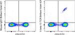
TCR V beta 13 Antibody (404-5797-82) in Flow
C57BL/6 mouse lymph node cells were stained with CD3 Monoclonal Antibody, FITC (Product # 11-0032-82) and 0.25 µg of Mouse IgG1 kappa Isotype Control, Brilliant Violet 421 (Product # 404-4714-81) (left) or 0.25 µg of TCR V beta 13 Monoclonal Antibody, Brilliant Violet 421 (right). Viable cells in the lymphocyte gate were used for analysis, as determined by 7-AAD (Product # 00-6993-50).
 in Flow")
产品信息
404-5797-82
产品规格
种属反应
Mouse
宿主/亚型
Mouse
/ IgG1, kappa
分类
Monoclonal
类型
Antibody
克隆号
MR12-3
偶联物
Brilliant Violet™ 421
Brilliant Violet™ 421
Brilliant Violet™ 421
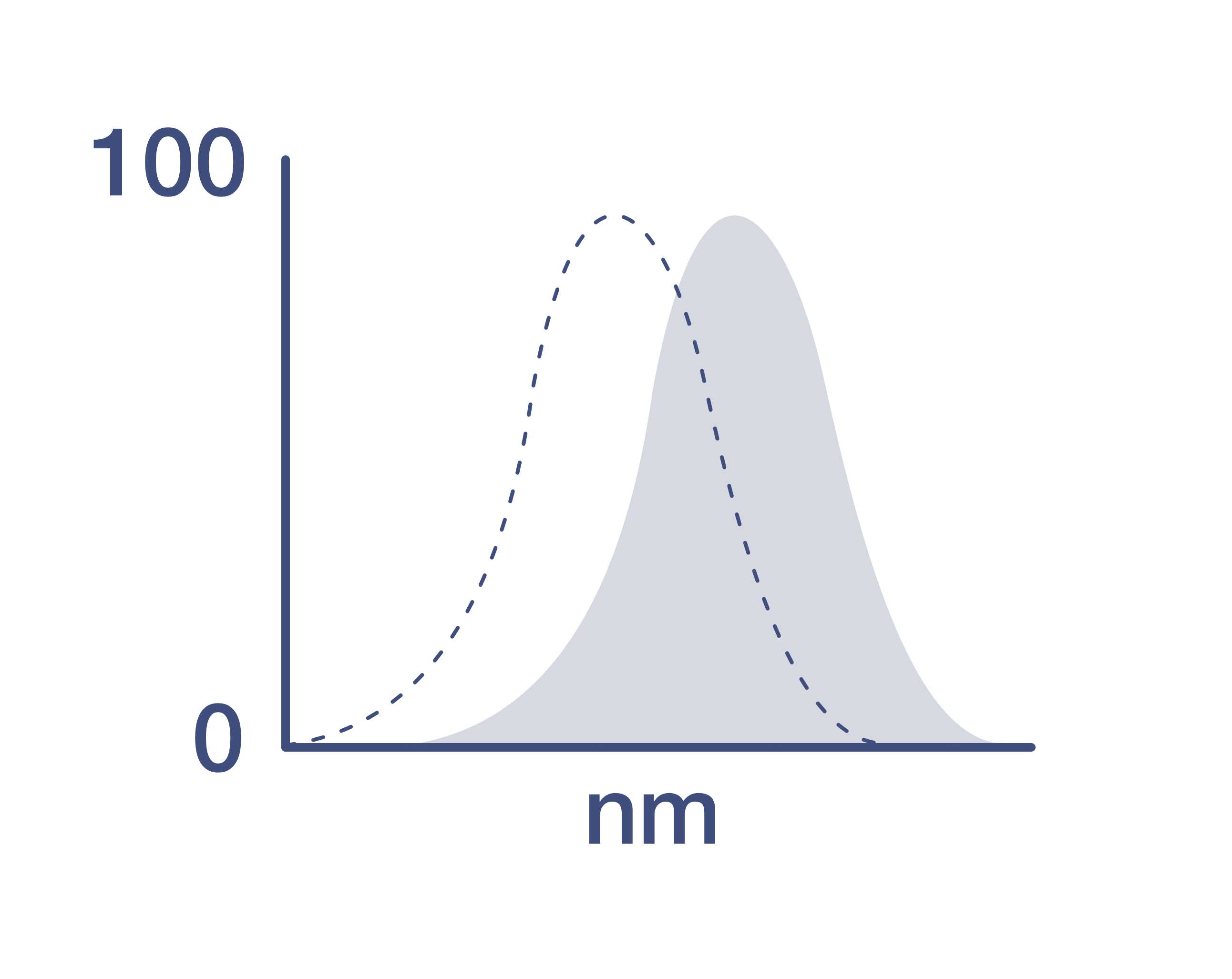
激发/发射光谱
406/423 nm
查看光谱
形式
liquid
浓度
0.2 mg/mL
规格
100 µg
纯化类型
Affinity chromatography
保存液
PBS, pH 7.2, with BSA
内含物
0.09% sodium azide
保存条件
4°C, store in dark, DO NOT FREEZE!
运输条件
Ambient (domestic); Wet ice (international)
RRID
产品详细信息
Description
This MR12-3 monoclonal antibody reacts with the T cell receptor (TCR) V beta 13 chain. Composed of an alpha and beta chain, TCR specificity is typically determined by Va, Ja, Vb, Db, and Jb gene rearrangement. The MR12-3 antibody recognizes the V beta 13 chains on T cells from mouse strains with the b haplotype of the Tcrb gene, including C57BL/6, B10, and C3H. V beta 13+ T cells are deleted in mouse strains with the a (e.g., SJL) and c (e.g., RIIIS/J) haplotypes. The MR12-3 monoclonal antibody has been reported to have blocking activity.
Applications Tested: This MR12-3 antibody has been tested by flow cytometric analysis of mouse lymph node cells. This may be used at less than or equal to 0.5 µg per test. A test is defined as the amount (µg) of antibody that will stain a cell sample in a final volume of 100 µL. Cell number should be determined empirically but can range from 10^5 to 10^8 cells/test. It is recommended that the antibody be carefully titrated for optimal performance in the assay of interest.
Blocking Buffers
When using two or more Super Bright, Brilliant Violet™, Brilliant Ultra Violet™, or other polymer dye-conjugated antibodies in a staining panel, it is recommended to use Super Bright Complete Staining Buffer (Product # SB-4401) or Brilliant Stain Buffer (Product # 00-4409-75) to minimize any non-specific polymer interactions. Please refer to the datasheet for Super Bright Staining Buffer or Brilliant Stain Buffer for more information.
Fixation
• Samples can be stored in IC Fixation Buffer (Product # 00-8222) (100 µL of cell sample + 100 µL of IC Fixation Buffer) or 1-step Fix/Lyse Solution (Product # 00-5333) for up to 3 days in the dark at 4°C with minimal impact on brightness and FRET efficiency/compensation.
• Some generalizations regarding fluorophore performance after fixation can be made, but clone specific performance should be determined empirically.
Excitation: 407 nm; Emission: 423 nm; Laser: Violet Laser.
BRILLIANT ULTRA VIOLET™ is a trademark or registered trademark of Becton, Dickinson and Company or its affiliates, and is used under license. Powered by Sirigen™.
靶标信息
The ability of T cell receptors (TCR) to discriminate foreign from self-peptides presented by major histocompatibility complex (MHC) class II molecules is essential for an effective adaptive immune response. TCR recognition of self-peptides has been linked to autoimmune disease. Mutant self-peptides have been associated with tumors. Engagement of TCRs by a family of bacterial toxins know as superantigens has been responsible for toxic shock syndrome. Autoantibodies to V beta segments of T cell receptors have been isolated from patients with rheumatoid arthritis (RA) and systemic lupus erythematosus (SLE). The autoantibodies block TH1-mediated inflammatory autodestructive reactions and are believed to be a method by which the immune system compensates for disease. Most human T cells express the TCR alpha-beta and either CD4 or CD8 molecule (single positive, SP). A small number of T cells lack both CD4 and CD8 (double negative, DN). Increased percentages of alpha-beta DN T cells have been identified in some autoimmune and immunodeficiency disorders. Gamma-delta T cells are primarily found within the epithelium. They show less TCR diversity and recognize antigens differently than alpha-beta T cells. Subsets of gamma-delta T cells have shown antitumor and immunoregulatory activity.
仅用于科研。不用于诊断过程。未经明确授权不得转售。
How to use the Panel Builder
Watch the video to learn how to use the Invitrogen Flow Cytometry Panel Builder to build your next flow cytometry panel in 5 easy steps.
篇参考文献 (0)
您是否在文献中引用过该产品?请点击下方按钮邮件告知我们。
生物信息学
蛋白别名: TCR V beta13; TCRV beta 13; TCRV beta13; Vb13; Vbeta13
Disclaimer

Clicking the images or links will redirect you to a website hosted by BenchSci that provides third-party scientific content. Neither the content nor the BenchSci technology and processes for selection have been evaluated by us; we are providing them as-is and without warranty of any kind, including for use or application of the Thermo Fisher Scientific products presented.

