Search
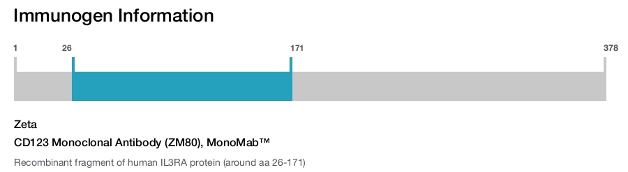
Zeta
CD123 Monoclonal Antibody (ZM80), MonoMab™
{{$productOrderCtrl.translations['antibody.pdp.commerceCard.promotion.promotions']}}
{{$productOrderCtrl.translations['antibody.pdp.commerceCard.promotion.viewpromo']}}
{{$productOrderCtrl.translations['antibody.pdp.commerceCard.promotion.promocode']}}: {{promo.promoCode}} {{promo.promoTitle}} {{promo.promoDescription}}. {{$productOrderCtrl.translations['antibody.pdp.commerceCard.promotion.learnmore']}}
Additional Information:
{{banner.description}}
产品信息
Z2390MS
产品规格
种属反应
Human
宿主/亚型
Mouse
/ IgG2b, kappa
分类
Monoclonal
类型
Antibody
克隆号
ZM80
抗原
Recombinant fragment of human IL3RA protein (around aa 26-171)
偶联物
Unconjugated
Unconjugated
Unconjugated
形式
Liquid
浓度
200 µg/mL
规格
100 µg
纯化类型
Protein A
保存液
tris with BSA, NP-40
内含物
<0.1% sodium azide
保存条件
4°C
运输条件
Ambient (domestic); Wet ice (international)
产品详细信息
A recommended positive control tissue for this product is Tonsil, however positive controls are not limited to this tissue type.
The primary antibody is intended for laboratory professional use in the detection of the corresponding protein in formalin-fixed, paraffin-embedded tissue stained in manual qualitative immunohistochemistry (IHC) testing. This antibody is intended to be used after the primary diagnosis of tumor has been made by conventional histopathology using non-immunological histochemical stains.
Human CD123, the 70 kd IL-3 receptor α chain (IL-3Rα), is associated with the 120-140 kd β subunit. The β chain is shared with the receptors for interleukins IL-5 and GM-CSF. IL-3Rα is expressed on hematopoietic progenitors and plays an important role in hematopoietic progenitor cell growth and differentiation. This antibody has been reported to block the binding of 125I-IL-3 to high and low affinity IL-3 receptors. In functional experiments, this antibody was found to inhibit acute myeloid leukemia cell proliferation, basophil histamine release, endothelial cell-mediated IL-8 secretion, and neutrophil transmigration. At the Fifth HLDA Workshop, the human IL-3 receptor was designated CD123. This antibody reacts with plasmacytoid dendritic cells (PDC). PDC plays a crucial role in the initiation of antiviral and antitumoral immune responses and produce type I interferons a and b. CD123 antibody is useful in diagnosis of Kikuchi disease, hyaline vascular type of Castleman disease, lupus, and primary cutaneous marginal zone B-cell lymphoma.
Antibody is used with formalin-fixed and paraffin-embedded sections. Pretreatment of deparaffinized tissue with heat-induced epitope retrieval or enzymatic retrieval is recommended. In general, immunohistochemical (IHC) staining techniques allow for the visualization of antigens via the sequential application of a specific antibody to the antigen (primary antibody), a secondary antibody to the primary antibody (link antibody), an enzyme complex and a chromogenic substrate with interposed washing steps. The enzymatic activation of the chromogen results in a visible reaction product at the antigen site. Results are interpreted using a light microscope and aid in the differential diagnosis of pathophysiological processes, which may or may not be associated with a particular antigen.
A positive tissue control must be run with every staining procedure performed. This tissue may contain both positive and negative staining cells or tissue components and serve as both the positive and negative control tissue. External Positive control materials should be fresh autopsy/biopsy/surgical specimens fixed, processed and embedded as soon as possible in the same manner as the patient sample (s). Positive tissue controls are indicative of correctly prepared tissues and proper staining methods. The tissues used for the external positive control materials should be selected from the patient specimens with well-characterized low levels of the positive target activity that gives weak positive staining. The low level of positivity for external positive controls is designed to ensure detection of subtle changes in the primary antibody sensitivity from instability or problems with the staining methodology. A tissue with weak positive staining is more suitable for optimal quality control and for detecting minor levels of reagent degradation.
Internal or external negative control tissue may be used depending on the guidelines and policies that govern the organization to which the end user belongs to. The variety of cell types present in many tissue sections offers internal negative control sites, but this should be verified by the user. The components that do not stain should demonstrate the absence of specific staining, and provide an indication of non-specific background staining. If specific staining occurs in the negative tissue control sites, results with the patient specimens must be considered invalid.
靶标信息
IL-3Ra (Interleukin-3 receptor subunit alpha) belongs to the type I cytokine receptor family and is a heterodimer with a unique alpha chain paired with the common beta (beta c or CDw131) subunit. IL-3Ra is responsible for the transmission of signal of Interleukin-3. IL-3Ra and IL-3Rb chain heterodimerization is induced by Interleukin-3, which is required for receptor activation but not high affinity binding. IL-3Ra is also reported that IL-3Ra subunits are overexpressed on acute myeloid leukemia (AML) blasts compared with normal hematopoietic cells and are thus potential targets for novel therapeutic agents.
仅用于科研。不用于诊断过程。未经明确授权不得转售。
篇参考文献 (0)
您是否在文献中引用过该产品?请点击下方按钮邮件告知我们。
生物信息学
蛋白别名: CD123; CD123 antigen; IL-3 receptor subunit alpha; IL-3R subunit alpha; interleukin 3 receptor, alpha (low affinity); Interleukin-3 receptor subunit alpha; membrane protein; unnamed protein product
基因别名: CD123; hIL-3Ra; IL-3R-alpha; IL3R; IL3RA; IL3RAY; IL3RX; IL3RY
UniProt ID: (Human) P26951
Entrez Gene ID: (Human) 3563
Disclaimer

Clicking the images or links will redirect you to a website hosted by BenchSci that provides third-party scientific content. Neither the content nor the BenchSci technology and processes for selection have been evaluated by us; we are providing them as-is and without warranty of any kind, including for use or application of the Thermo Fisher Scientific products presented.

