Search
Invitrogen
FceR1 alpha Monoclonal Antibody (AER-37 (CRA1)), Brilliant Violet™ 786, eBioscience™
{{$productOrderCtrl.translations['antibody.pdp.commerceCard.promotion.promotions']}}
{{$productOrderCtrl.translations['antibody.pdp.commerceCard.promotion.viewpromo']}}
{{$productOrderCtrl.translations['antibody.pdp.commerceCard.promotion.promocode']}}: {{promo.promoCode}} {{promo.promoTitle}} {{promo.promoDescription}}. {{$productOrderCtrl.translations['antibody.pdp.commerceCard.promotion.learnmore']}}
Additional Information:
{{banner.description}}
")
图: 1 / 1
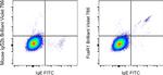
FceR1 alpha Antibody (417-5899-42) in Flow
Normal human peripheral blood cells were stained with IgE Monoclonal Antibody, FITC (Product # 11-6986-42) and Mouse IgG2b kappa Isotype Control, Brilliant Violet 786 (Product # 417-4732-81) (left) or FceR1 alpha Monoclonal Antibody, Brilliant Violet 786 (right). Viable cells in the lymphocyte gate were used for analysis, as determined by 7-AAD (Product # 00-6993-50) (note: basophils reside in FCS/SSC position of lymphocytes).
Please note: We are reviewing Western blot images included in the antibody testing data in our catalog, including those provided by third parties. Unless expressly labeled or annotated as “raw-unedited”, Western blot images included in the antibody testing data in our catalog may have been edited, optimized or otherwise adjusted for presentation.
 in Flow")
产品信息
417-5899-42
产品规格
种属反应
Human
宿主/亚型
Mouse
/ IgG2b, kappa
分类
Monoclonal
类型
Antibody
克隆号
AER-37 (CRA1)
偶联物
Brilliant Violet™ 786
Brilliant Violet™ 786
Brilliant Violet™ 786
激发/发射光谱
406/788 nm
查看光谱
形式
Liquid
浓度
5 µL/Test
纯化类型
Affinity chromatography
保存液
PBS, pH 7.2, with BSA
内含物
0.09% sodium azide
保存条件
4°C, store in dark
运输条件
Ambient (domestic); Wet ice (international)
RRID
产品详细信息
Description
The AER-37 monoclonal antibody reacts with the Fc epsilon RI alpha subunit, an IgE-binding subunit lacking signal-transducing ability.
Applications Tested
This AER-37 (CRA1) antibody has been pre-diluted and tested by flow cytometric analysis of normal human peripheral blood cells. This may be used at 5 µL (0.5 µg) per test. A test is defined as the amount (µg) of antibody that will stain a cell sample in a final volume of 100 µL. Cell number should be determined empirically but can range from 10^5 to 10^8 cells/test.
Blocking Buffers
When using two or more Super Bright, Brilliant Violet™, Brilliant Ultra Violet™, or other polymer dye-conjugated antibodies in a staining panel, it is recommended to use Super Bright Complete Staining Buffer (Product # SB-4401) or Brilliant Stain Buffer (Product # 00-4409-75) to minimize any non-specific polymer interactions. Please refer to the datasheet for Super Bright Staining Buffer or Brilliant Stain Buffer for more information.
Light sensitivity
This tandem dye is sensitive to photo-induced oxidation. Please protect this vial and stained samples from light.
Fixation
• Samples can be stored in IC Fixation Buffer (Product # 00-8222) (100 µL of cell sample + 100 µL of IC Fixation Buffer) or 1-step Fix/Lyse Solution (Product # 00-5333) for up to 3 days in the dark at 4°C with minimal impact on brightness and FRET efficiency/compensation.
• Some generalizations regarding fluorophore performance after fixation can be made, but clone specific performance should be determined empirically.
• Our internal testing suggests that Brilliant Violet™ 786 (BV786) is not compatible with methanol-based fixation.
Excitation: 407 nm; Emission: 786 nm; Laser: Violet Laser.
BRILLIANT ULTRA VIOLET™ is a trademark or registered trademark of Becton, Dickinson and Company or its affiliates, and is used under license. Powered by Sirigen™.
靶标信息
Fc epsilon RI alpha, also known as FceR1 alpha, is a subunit of the high-affinity receptor for IgE, primarily expressed on mast cells and basophils. Its expression is upregulated by the presence of IgE. Fc epsilon RI alpha forms a tetrameric complex with one beta and two gamma subunits, which are essential for signal transduction. The beta and gamma subunits contain immunoreceptor tyrosine-based activation motifs (ITAMs). The Fc epsilon RI complex plays a crucial role in triggering IgE-mediated allergic reactions. When allergen-bound IgE molecules bring together two or more high-affinity IgE receptors, it leads to the release of mediators such as histamine, which are responsible for allergy symptoms. This process couples allergens and mast cells to initiate inflammatory responses characteristic of allergic disorders like hay fever and asthma. The release of histamine and proteases also leads to the synthesis of prostaglandins and leukotrienes, which are potent effectors of the hypersensitivity response.
仅用于科研。不用于诊断过程。未经明确授权不得转售。
How to use the Panel Builder
Watch the video to learn how to use the Invitrogen Flow Cytometry Panel Builder to build your next flow cytometry panel in 5 easy steps.
篇参考文献 (0)
您是否在文献中引用过该产品?请点击下方按钮邮件告知我们。
生物信息学
蛋白别名: fc epsilon r1; Fc-epsilon RI-alpha; FcERI; High affinity immunoglobulin epsilon receptor subunit alpha; IgE Fc receptor subunit alpha; RP11-550P17.3
基因别名: FCE1A; FCER1A
Entrez Gene ID: (Human) 2205
Disclaimer

Clicking the images or links will redirect you to a website hosted by BenchSci that provides third-party scientific content. Neither the content nor the BenchSci technology and processes for selection have been evaluated by us; we are providing them as-is and without warranty of any kind, including for use or application of the Thermo Fisher Scientific products presented.

