Search
Invitrogen
HCoV-229E Spike S2 Recombinant Rabbit Monoclonal Antibody (6P2H6)
{{$productOrderCtrl.translations['antibody.pdp.commerceCard.promotion.promotions']}}
{{$productOrderCtrl.translations['antibody.pdp.commerceCard.promotion.promocode']}}: {{promo.promoCode}} {{promo.promoTitle}} {{promo.promoDescription}}. {{$productOrderCtrl.translations['antibody.pdp.commerceCard.promotion.learnmore']}}
{{$ctrl.translations['antibody.pdp.skuNotes.additionalInformation']}}:
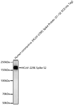
图: 1 / 1
HCoV-229E Spike S2 Antibody (MA575570) in WB
Western blot analysis of HCoV-229E Spike S2 in Human coronavirus (HCoV-229E) Spike Protein (S1+S2 ECD His Tag) (100 ng per lane). Samples were blocked with 3% nonfat dry milk in TBST, incubated with monoclonal HCoV-229E Spike S2 (Product # MA575570) antibody at a dilution of 1:3,000 (overnight for 1.5h at RT), followed by HRP-conjugated Goat anti-Rabbit IgG (H+L) (1:10,000) and ECL. Exposure time: 45s.
Please note: We are reviewing Western blot images included in the antibody testing data in our catalog, including those provided by third parties. Unless expressly labeled or annotated as “raw-unedited”, Western blot images included in the antibody testing data in our catalog may have been edited, optimized or otherwise adjusted for presentation.

产品信息
MA575570
产品规格
种属反应
Virus
宿主/亚型
Rabbit
/ IgG
Expression System
HEK293 cells
分类
Recombinant Monoclonal
类型
Antibody
克隆号
6P2H6
抗原
Recombinant protein (or fragment).This information is considered to be commercially sensitive.
偶联物
Unconjugated
Unconjugated
Unconjugated
形式
Liquid
浓度
0.61 mg/mL
规格
61 µg
纯化类型
Affinity chromatography
保存液
PBS, pH 7.4, with 0.05% BSA, 50% glycerol
内含物
0.05% ProClin 300
保存条件
-20°C, Avoid Freeze/Thaw Cycles
运输条件
Ambient (domestic); Wet ice (international)
靶标信息
S1 region attaches the virion to the cell membrane by interacting with host ANPEP/aminopeptidase N, initiating the infection. Binding to the receptor probably induces conformational changes in the S glycoprotein unmasking the fusion peptide of S2 region and activating membranes fusion. S2 region belongs to the class I viral fusion protein. Under the current model, the protein has at least 3 conformational states: pre-fusion native state, pre-hairpin intermediate state, and post-fusion hairpin state. During viral and target cell membrane fusion, the coiled coil regions (heptad repeats) regions assume a trimer-of-hairpins structure, positioning the fusion peptide in close proximity to the C-terminal region of the ectodomain. The formation of this structure appears to drive apposition and subsequent fusion of viral and target cell membranes. [Uniprot]
仅用于科研。不用于诊断过程。未经明确授权不得转售。
篇参考文献 (0)
您是否在文献中引用过该产品?请点击下方按钮邮件告知我们。
生物信息学
蛋白别名: 229E; Coronavirus; HCoV; S2; Spike
Disclaimer

Clicking the images or links will redirect you to a website hosted by BenchSci that provides third-party scientific content. Neither the content nor the BenchSci technology and processes for selection have been evaluated by us; we are providing them as-is and without warranty of any kind, including for use or application of the Thermo Fisher Scientific products presented.

