Search
Invitrogen
Lucifer Yellow Polyclonal Antibody, Biotin
{{$productOrderCtrl.translations['antibody.pdp.commerceCard.promotion.promotions']}}
{{$productOrderCtrl.translations['antibody.pdp.commerceCard.promotion.promocode']}}: {{promo.promoCode}} {{promo.promoTitle}} {{promo.promoDescription}}. {{$productOrderCtrl.translations['antibody.pdp.commerceCard.promotion.learnmore']}}
{{$ctrl.translations['antibody.pdp.skuNotes.additionalInformation']}}:
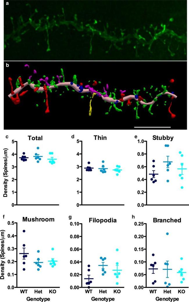
图: 1 / 5
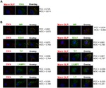
Lucifer Yellow Antibody (A-5751) in ICC/IF
Molecular autism 2020 -
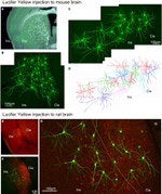
Spine density and morphology were similar in WT, Shank3 -Het, and Shank3 -KO. a Representative example of a deconvolved confocal image of a basal dendritic segment and b its 3D reconstruction, with the dendritic segment shown in light pink, thin spines in green, stubby in blue, mushroom in red, filopodia in yellow, and branched spines in magenta. Scale bar = 5 um. c Total spine density and density by spine ... View More
Please note: We are reviewing Western blot images included in the antibody testing data in our catalog, including those provided by third parties. Unless expressly labeled or annotated as “raw-unedited”, Western blot images included in the antibody testing data in our catalog may have been edited, optimized or otherwise adjusted for presentation.


产品信息
A-5751
应用
建议稀释比
已发表文章
产品规格
种属反应
Chemical
已发表种属
Chemical
宿主/亚型
Rabbit
/ IgG
分类
Polyclonal
类型
Antibody
抗原
Lucifer Yellow
偶联物
Biotin
Biotin
Biotin
形式
Liquid
浓度
3 mg/mL
规格
1.5 mg
纯化类型
IgG fraction
保存液
PBS, pH 7.2
内含物
5mM sodium azide
保存条件
4°C
运输条件
Ambient (domestic); Wet ice (international)
RRID
靶标信息
Even though the cell soma of a lucifer yellow-filled neuron may be brightly stained, its finer processes can sometimes be faint and may fade rapidly or be obscured by the more intensely stained portions of the neuron. By using anti-lucifer yellow dye antibodies in conjunction with standard enzyme-mediated immunohistochemical methods one can develop a more permanent, fade-free signal for light microscopy.
仅用于科研。不用于诊断过程。未经明确授权不得转售。
生物信息学
蛋白别名: Lucifer Yellow Dye
Disclaimer

Clicking the images or links will redirect you to a website hosted by BenchSci that provides third-party scientific content. Neither the content nor the BenchSci technology and processes for selection have been evaluated by us; we are providing them as-is and without warranty of any kind, including for use or application of the Thermo Fisher Scientific products presented.

