Search
Invitrogen
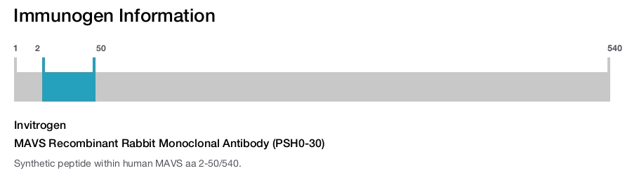
MAVS Recombinant Rabbit Monoclonal Antibody (PSH0-30)
{{$productOrderCtrl.translations['antibody.pdp.commerceCard.promotion.promotions']}}
{{$productOrderCtrl.translations['antibody.pdp.commerceCard.promotion.viewpromo']}}
{{$productOrderCtrl.translations['antibody.pdp.commerceCard.promotion.promocode']}}: {{promo.promoCode}} {{promo.promoTitle}} {{promo.promoDescription}}. {{$productOrderCtrl.translations['antibody.pdp.commerceCard.promotion.learnmore']}}
Additional Information:
{{banner.description}}
")
图: 1 / 4
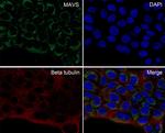
MAVS Antibody (MA5-50801) in ICC/IF
Immunocytochemistry analysis of MAVS in A431 cells. Cells were fixed in 4% paraformaldehyde for 10 minutes at 37 &(8)451;, permeabilized with 0.05% Triton X-100 in PBS for 20 minutes, and then blocked with 2% negative goat serum for 30 minutes at room temperature. Cells were incubated with MAVS recombinant monoclonal antibody (Product # MA5-50801) with a dilution of 1:100 in 2% negative goat serum overnight at 4 &(8)451;. Followed by secondary antibody Goat Anti-Rabbit IgG H&L (Alexa Fluor® 488) at a dilution of 1:1,000. Nuclear DNA was la... View More
 in ICC/IF")
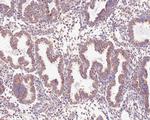
 in IHC (P)")
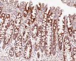
 in IHC (P)")
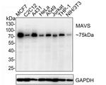
 in WB")
产品信息
MA5-50801
产品规格
宿主/亚型
Rabbit
/ IgG
Expression System
HEK293 cells
分类
Recombinant Monoclonal
类型
Antibody
克隆号
PSH0-30
抗原
Synthetic peptide within human MAVS aa 2-50/540.
偶联物
Unconjugated
Unconjugated
Unconjugated
形式
Liquid
浓度
1 mg/mL
规格
100 µg
保存条件
Store at 4°C short term. For long term storage, store at -20°C, avoiding freeze/thaw cycles.
运输条件
Wet ice
RRID
产品详细信息
Positive control: MCF7 cell lysate, C2C12 cell lysate, human colon tissue, human endometrium tissue, A431 cell.
Predicted band size: 56 kDa
Subcellular Location: Membrane, Mitochondrion, Mitochondrion outer membrane, Peroxisome.
靶标信息
Two distinct signaling pathways activate the host innate immunity against viral infection. One pathway is reliant on members of the Toll-like receptor (TLR) family while the other uses the RNA helicase RIG-I as a receptor for intracellular viral double-stranded RNA as a trigger for the immune response. MAVS is a mitochondrial membrane protein that was identified as a critical component in the IFN beta signaling pathways that recruits IRF-3 to RIG-I, leading to its activation and that of NF-kappa-B. MAVS is also thought to interact with other components of the innate immune pathway such as the TLR adapter protein TRIF, TRAF2 and TRAF6. MAVS also interacts with the IKK-alpha, IKK-beta and IKK-iota kinases through its C-terminal region. Cleavage of this region by the Hepatitis C virus (HCV) protease allows HCV to escape the host immune system. Multiple isoforms of MAVS are known to exist.
仅用于科研。不用于诊断过程。未经明确授权不得转售。
篇参考文献 (0)
您是否在文献中引用过该产品?请点击下方按钮邮件告知我们。
Disclaimer

Clicking the images or links will redirect you to a website hosted by BenchSci that provides third-party scientific content. Neither the content nor the BenchSci technology and processes for selection have been evaluated by us; we are providing them as-is and without warranty of any kind, including for use or application of the Thermo Fisher Scientific products presented.

