Search
Invitrogen
Phospho-CD247 (CD3 zeta) (Tyr142) Recombinant Mouse Monoclonal Antibody (3ZBR4S), Alexa Fluor™ Plus 647
{{$productOrderCtrl.translations['antibody.pdp.commerceCard.promotion.promotions']}}
{{$productOrderCtrl.translations['antibody.pdp.commerceCard.promotion.promocode']}}: {{promo.promoCode}} {{promo.promoTitle}} {{promo.promoDescription}}. {{$productOrderCtrl.translations['antibody.pdp.commerceCard.promotion.learnmore']}}
{{$ctrl.translations['antibody.pdp.skuNotes.additionalInformation']}}:
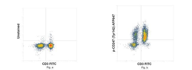
图: 1 / 3
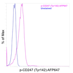
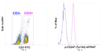
Phospho-CD247 (CD3 zeta) (Tyr142) Antibody (740251MP647) in Flow
Flow cytometry analysis of Phospho-CD247 (CD3 zeta) (Tyr142) Recombinant Mouse Monoclonal Antibody (3ZBR4S), Alexa Fluor™ Plus 647 (Product # 740251MP647) was performed using Human PBMCs stimulated with CD3 and CD28 for 15 min. Cells were fixed with eBioscience™ IC Fixation buffer (Product # 00-8222-49), permeabilized and stained with CD3 Monoclonal Antibody (UCHT1) FITC, eBioscience™ (Product # 11-0038-42)... View More
Please note: We are reviewing Western blot images included in the antibody testing data in our catalog, including those provided by third parties. Unless expressly labeled or annotated as “raw-unedited”, Western blot images included in the antibody testing data in our catalog may have been edited, optimized or otherwise adjusted for presentation.


产品信息
740251MP647
产品规格
种属反应
Human
宿主/亚型
Mouse
/ IgG2b, kappa
Expression System
Expi293
分类
Recombinant Monoclonal
类型
Antibody
克隆号
3ZBR4S
偶联物
Alexa Fluor™ Plus 647
Alexa Fluor™ Plus 647
Alexa Fluor™ Plus 647
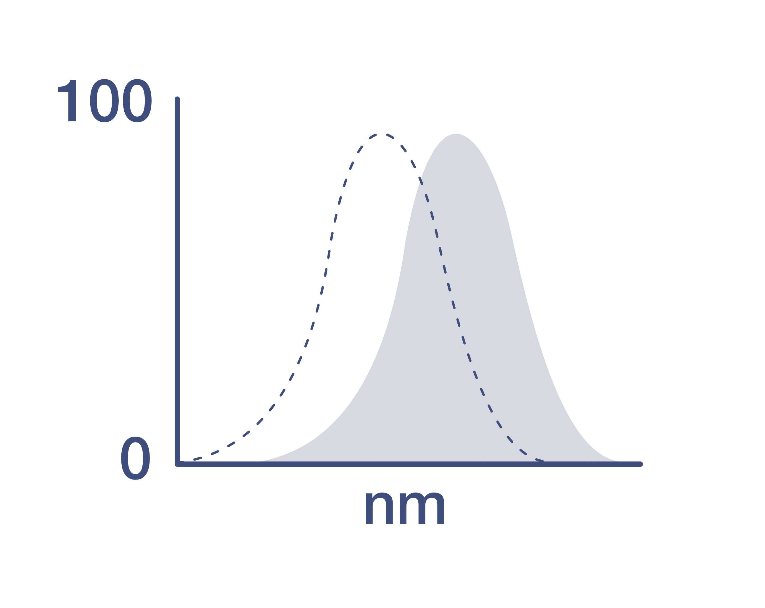
激发/发射光谱
658/675 nm
查看光谱
形式
Liquid
浓度
1 mg/mL
规格
50 µg
纯化类型
Protein A/G
保存液
proprietary buffer, pH 6.3-7.3
内含物
0.008% Bromonitrodioxane, 0.008% Methylisothiazolone
保存条件
4°C, store in dark, DO NOT FREEZE!
运输条件
Wet ice
产品详细信息
Alexa Fluor™ Plus recombinant antibodies are conjugated using new, proprietary dye chemistry so you can generate stunning data. Alexa Fluor™ Plus antibodies represent an advancement in fluorescent conjugate technology. Alexa Fluor™ Plus antibodies provide brighter signal compared to leading Alexa Fluor™ antibodies, providing you with better signal-to-noise for your critical experiments. These antibodies show better specificity and lot-to-lot consistency as these are recombinant antibodies, generated by cloning specific genes for the desired antibodies into an expression vector and expressed in vitro.
Using conjugate solutions: Centrifuge the protein conjugate solution briefly in a microcentrifuge before use; add only the supernatant to the experiment. This step will help eliminate any protein aggregates that may have formed during storage, thereby reducing nonspecific background staining.
It is recommended that the antibody be carefully titrated for optimal performance in the assay of interest.
Excitation: 658 nm; Emission: 675 nm; Laser: Red Laser
Filtration: 0.2 µm post-manufacturing filtered.
靶标信息
CD3z complex is crucial in transducing antigen-recognition signals into the cytoplasm of T cells and in regulating the cell surface expression of the TCR complex. T cell activation through the antigen receptor (TCR) involves the cytoplasmic tails of the CD3 subunits CD3 gamma, CD3 delta, CD3 epsilon and CD3 zeta. These CD3 subunits are structurally related members of the immunoglobulins super family encoded by closely linked genes on human chromosome 11. The CD3 components have long cytoplasmic tails that associate with cytoplasmic signal transduction molecules, and this association is mediated at least in part by a double tyrosine-based motif present in a single copy in the CD3 subunits. CD3 may play a role in TCR-induced growth arrest, cell survival and proliferation. The CD3 antigen is present on 68-82% of normal peripheral blood lymphocytes, 65-85% of thymocytes and Purkinje cells in the cerebellum. It is never expressed on B or NK cells. Decreased percentages of T lymphocytes may be observed in some autoimmune diseases. Diseases associated with CD3z include Immunodeficiency 25 and T-B+ Severe Combined Immunodeficiency Due To Cd3delta/Cd3epsilon/Cd3zeta.
仅用于科研。不用于诊断过程。未经明确授权不得转售。
篇参考文献 (0)
您是否在文献中引用过该产品?请点击下方按钮邮件告知我们。
生物信息学
蛋白别名: CD247; CD247 antigen, zeta subunit; CD3-epsilon; CD3e antigen; CD3Z antigen, zeta polypeptide (TiT3 complex); CD3zeta chain; component of the TCR/CD3 complex; FLJ18683; T-cell antigen receptor complex, zeta subunit of CD3; T-cell receptor T3 zeta chain; T3E; TCR zeta; TCR zeta chain; TCRE; tissue factor-targeting CAR1 dimer; tissue factor-targeting CAR1 monomer; unnamed protein product
基因别名: CD3-ZETA; CD3H; CD3Q; CD3Z; CD3ZETA; IMD25; T3Z; TCRZ
UniProt ID: (Human) P20963
Entrez Gene ID: (Human) 919
Disclaimer

Clicking the images or links will redirect you to a website hosted by BenchSci that provides third-party scientific content. Neither the content nor the BenchSci technology and processes for selection have been evaluated by us; we are providing them as-is and without warranty of any kind, including for use or application of the Thermo Fisher Scientific products presented.

