Search
Invitrogen
Phospho-STAT3 (Tyr705) Recombinant Mouse Monoclonal Antibody (LUVNKLA), Alexa Fluor™ Plus 647
{{$productOrderCtrl.translations['antibody.pdp.commerceCard.promotion.promotions']}}
{{$productOrderCtrl.translations['antibody.pdp.commerceCard.promotion.viewpromo']}}
{{$productOrderCtrl.translations['antibody.pdp.commerceCard.promotion.promocode']}}: {{promo.promoCode}} {{promo.promoTitle}} {{promo.promoDescription}}. {{$productOrderCtrl.translations['antibody.pdp.commerceCard.promotion.learnmore']}}
Additional Information:
{{banner.description}}
 Antibody in Flow Cytometry (Flow)")
图: 1 / 2
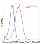
Phospho-STAT3 (Tyr705) Antibody (740067MP647) in Flow
Flow cytometry analysis of Phospho-STAT3 was performed using Jurkat cells and Jurkat cells treated with 150 ng/mL human interferon alpha for 30 minutes. Cells were fixed with IC Fixation Buffer (Product # 00-8222-49), permeabilized with ice-cold methanol and stained intracellularly with 0.25 µg of Phospho-STAT3 (Tyr705) Recombinant Mouse Monoclonal Antibody (LUVNKLA), Alexa Fluor™ Plus 647 (Product # 740067... View More

Please note: We are reviewing Western blot images included in the antibody testing data in our catalog, including those provided by third parties. Unless expressly labeled or annotated as “raw-unedited”, Western blot images included in the antibody testing data in our catalog may have been edited, optimized or otherwise adjusted for presentation.
 Antibody (740067MP647) in Flow")
 Antibody (740067MP647)")
产品信息
740067MP647
产品规格
种属反应
Human,
Mouse
宿主/亚型
Mouse
/ IgG2b, kappa
Expression System
Expi293
分类
Recombinant Monoclonal
类型
Antibody
克隆号
LUVNKLA
偶联物
Alexa Fluor™ Plus 647
Alexa Fluor™ Plus 647
Alexa Fluor™ Plus 647
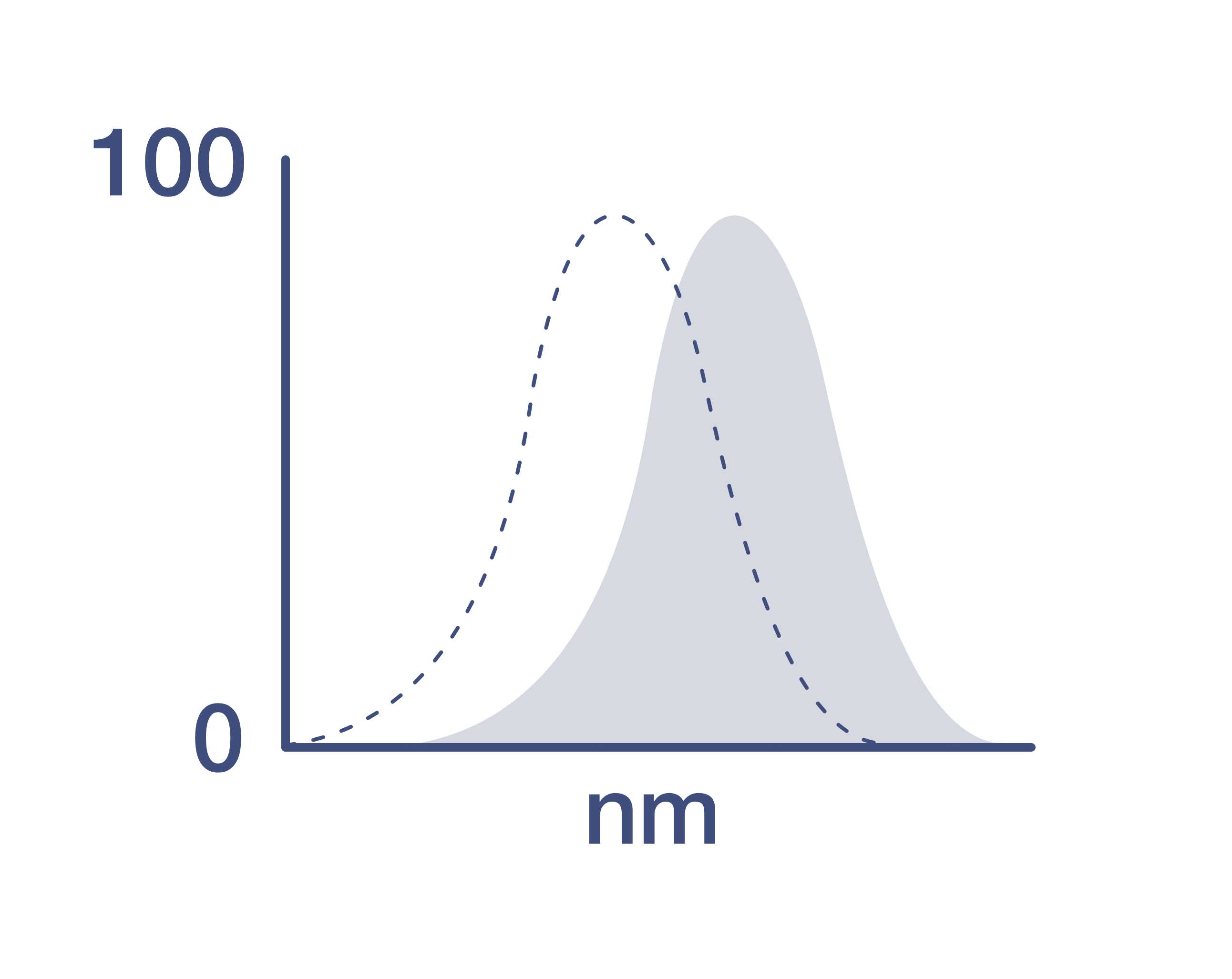
激发/发射光谱
658/675 nm
查看光谱
形式
Liquid
浓度
1.0 mg/mL
规格
50 µg
纯化类型
Protein A/G
保存液
proprietary buffer, pH 6.8
内含物
0.008% Bromonitrodioxane, 0.008% Methylisothiazolone
保存条件
4°C, store in dark, DO NOT FREEZE!
运输条件
Wet ice
RRID
产品详细信息
Alexa Fluor™ Plus recombinant antibodies are conjugated using new, proprietary dye chemistry so you can generate stunning data. Alexa Fluor™ Plus antibodies represent an advancement in fluorescent conjugate technology. Alexa Fluor™ Plus antibodies provide brighter signal compared to leading Alexa Fluor™ antibodies, providing you with better signal-to-noise for your critical experiments. These antibodies show better specificity and lot-to-lot consistency as these are recombinant antibodies, generated by cloning specific genes for the desired antibodies into an expression vector and expressed in vitro.
Using conjugate solutions: Centrifuge the protein conjugate solution briefly in a microcentrifuge before use; add only the supernatant to the experiment. This step will help eliminate any protein aggregates that may have formed during storage, thereby reducing nonspecific background staining.
Applications Tested: This LUVNKLA antibody has been tested by flow cytometric analysis of Jurkat cells. This may be used for flow cytometry at 0.25 µg per test. A test is defined as the amount (µg) of antibody that will stain a cell sample in a final volume of 100 µL. Cell number should be determined empirically but can range from 10^5 to 10^8 cells/test. It is recommended that the antibody be carefully titrated for optimal performance in the assay of interest.
Excitation: 658 nm; Emission: 675 nm; Laser: Red Laser
Filtration: 0.2 µm post-manufacturing filtered.
靶标信息
STAT3 belongs to the family of STAT (signal transducers and activators of transcription) proteins which are phosphorylated by receptor associated kinases, translocate to the nucleus, and act as transcription factors in response to cytokines and growth factors. Coactivators such as CREB-binding protein are required for the transcriptional activation by STAT3. STAT3 can also be activated by Interferon-alpha, Interferon-gamma, EGF, PDGF and IL6. Phosphorylation on tyrosine 705 by JAK1 and JAK2 is essential for STAT3 dimer formation, nuclear translocation, and DNA binding activity. In humans, the STAT3 gene is located on the q arm of chromosome 17. STAT3 has been shown to be activated by IFN-alpha but not IFN-beta. The transcription factors associated with STAT3 are c-Jun and cyclic AMP-responsive enhancer binding protein (CREB). STAT3 mediates the expression of a variety of genes in response to cell stimuli, and thus plays a key role in many cellular processes such as cell growth and apoptosis. The small GTPase Rac1 has been shown to bind and regulate the activity of STAT3 while the PIAS3 protein is a specific inhibitor of STAT3. Three alternatively spliced transcript variants encoding distinct isoforms of STAT3 have been described. Deletion of the STAT3 gene in knock-out mice was lethal at the early embryonic stage.
仅用于科研。不用于诊断过程。未经明确授权不得转售。
篇参考文献 (0)
您是否在文献中引用过该产品?请点击下方按钮邮件告知我们。
生物信息学
蛋白别名: acute phase response factor; acute-phase response factor; DNA-binding protein APRF; FLJ20882; MGC16063; STAT; Stat3; transcription factor; unnamed protein product
基因别名: 1110034C02Rik; ADMIO; ADMIO1; APRF; HIES
UniProt ID: (Human) P40763, (Mouse) P42227
Entrez Gene ID: (Human) 6774, (Mouse) 20848
transcription regulatory region sequence-specific DNA binding
RNA polymerase II core promoter proximal region sequence-specific DNA binding
RNA polymerase II transcription factor activity, sequence-specific DNA binding
transcriptional activator activity, RNA polymerase II transcription regulatory region sequence-specific binding
DNA binding
transcription factor activity, sequence-specific DNA binding
RNA binding
RNA polymerase II transcription factor activity, ligand-activated sequence-specific DNA binding
receptor binding
protein binding
protein kinase binding
protein phosphatase binding
chromatin DNA binding
CCR5 chemokine receptor binding
glucocorticoid receptor binding
signaling adaptor activity
identical protein binding
protein homodimerization activity
sequence-specific DNA binding
protein dimerization activity
RNA polymerase II sequence-specific DNA binding transcription factor binding
primary miRNA binding
lncRNA binding
DNA-binding transcription factor binding
protein sequestering activity
RNA sequestering activity
acetylation-dependent protein binding
temperature homeostasis
eye photoreceptor cell differentiation
regulation of cytokine production
plasma cell differentiation
response to ischemia
regulation of transcription, DNA-templated
regulation of transcription from RNA polymerase II promoter
transcription from RNA polymerase II promoter
protein import into nucleus
defense response
inflammatory response
signal transduction
transforming growth factor beta receptor signaling pathway
I-kappaB kinase/NF-kappaB signaling
JAK-STAT cascade
cell proliferation
positive regulation of cell proliferation
negative regulation of cell proliferation
gene expression
negative regulation of autophagy
positive regulation of vascular endothelial growth factor production
positive regulation of gene expression
negative regulation of gene expression
negative regulation of hydrogen peroxide biosynthetic process
cytokine-mediated signaling pathway
stem cell population maintenance
signal transduction involved in regulation of gene expression
cell differentiation
positive regulation of cell migration
intracellular receptor signaling pathway
endoplasmic reticulum unfolded protein response
response to estradiol
positive regulation of interleukin-1 beta production
positive regulation of interleukin-10 production
positive regulation of interleukin-6 production
positive regulation of interleukin-8 production
positive regulation of tumor necrosis factor production
cellular response to hormone stimulus
leptin-mediated signaling pathway
response to cytokine
interleukin-15-mediated signaling pathway
interleukin-2-mediated signaling pathway
interleukin-9-mediated signaling pathway
interleukin-11-mediated signaling pathway
interleukin-23-mediated signaling pathway
oncostatin-M-mediated signaling pathway
regulation of multicellular organism growth
regulation of cell proliferation
glucose homeostasis
eating behavior
mRNA transcription from RNA polymerase II promoter
regulation of I-kappaB kinase/NF-kappaB signaling
positive regulation of I-kappaB kinase/NF-kappaB signaling
negative regulation of I-kappaB kinase/NF-kappaB signaling
response to peptide hormone
protein kinase B signaling
negative regulation of neuron apoptotic process
cellular response to leptin stimulus
response to leptin
positive regulation of erythrocyte differentiation
positive regulation of Notch signaling pathway
positive regulation of angiogenesis
negative regulation of glycolytic process
positive regulation of transcription, DNA-templated
positive regulation of transcription from RNA polymerase II promoter
regulation of mitochondrial membrane permeability
astrocyte differentiation
leukemia inhibitory factor signaling pathway
negative regulation of inflammatory response
positive regulation of inflammatory response
positive regulation of phagocytosis
modulation of synaptic transmission
positive regulation of multicellular organismal process
regulation of cell cycle
radial glial cell differentiation
regulation of feeding behavior
growth hormone receptor signaling pathway
JAK-STAT cascade involved in growth hormone signaling pathway
interleukin-6-mediated signaling pathway
ciliary neurotrophic factor-mediated signaling pathway
cellular response to cytokine stimulus
T-helper 17 type immune response
T-helper 17 cell lineage commitment
positive regulation of extracellular matrix disassembly
inflammatory response to wounding
energy homeostasis
cellular response to interleukin-17
receptor signaling pathway via STAT
postsynapse to nucleus signaling pathway
negative regulation of inflammatory response to wounding
interleukin-10-mediated signaling pathway
negative regulation of cytokine production involved in inflammatory response
positive regulation of cytokine production involved in inflammatory response
regulation of cellular response to hypoxia
positive regulation of growth factor dependent skeletal muscle satellite cell proliferation
positive regulation of pri-miRNA transcription from RNA polymerase II promoter
regulation of ATP metabolic process
positive regulation of vascular endothelial cell proliferation
negative regulation of 3'-UTR-mediated mRNA stabilization
negative regulation of primary miRNA processing
negative regulation of stem cell differentiation
positive regulation of ATP biosynthetic process
negative regulation of neuron migration
negative regulation of intrinsic apoptotic signaling pathway
negative regulation of transcription from RNA polymerase II promoter
response to hypoxia
acute-phase response
immune response
nervous system development
response to ethanol
Disclaimer

Clicking the images or links will redirect you to a website hosted by BenchSci that provides third-party scientific content. Neither the content nor the BenchSci technology and processes for selection have been evaluated by us; we are providing them as-is and without warranty of any kind, including for use or application of the Thermo Fisher Scientific products presented.

