Search
Disclaimer

Clicking the images or links will redirect you to a website hosted by BenchSci that provides third-party scientific content. Neither the content nor the BenchSci technology and processes for selection have been evaluated by us; we are providing them as-is and without warranty of any kind, including for use or application of the Thermo Fisher Scientific products presented.
Invitrogen
Triadin Monoclonal Antibody (IIG12)
{{$productOrderCtrl.translations['antibody.pdp.commerceCard.promotion.promotions']}}
{{$productOrderCtrl.translations['antibody.pdp.commerceCard.promotion.viewpromo']}}
{{$productOrderCtrl.translations['antibody.pdp.commerceCard.promotion.promocode']}}: {{promo.promoCode}} {{promo.promoTitle}} {{promo.promoDescription}}. {{$productOrderCtrl.translations['antibody.pdp.commerceCard.promotion.learnmore']}}
")
 in WB")
产品信息
MA3-931
应用
建议稀释比
已发表文章
产品规格
已发表种属
Mouse,
Rabbit
宿主/亚型
Mouse
/ IgG1
分类
Monoclonal
类型
Antibody
克隆号
IIG12
抗原
Rabbit skeletal muscle triads.
偶联物
形式
Liquid
浓度
Conc. Not Determined
保存条件
-20°C, Avoid Freeze/Thaw Cycles
运输条件
Wet ice
RRID
产品详细信息
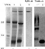
MA3-931 detects triadin from mouse and rabbit skeletal muscle tissues. This antibody does not detect triadin from cardiac tissues.
MA3-931 has been successfully used in Western blot and immunohistochemistry procedures. By Western blot, this antibody detects a single ~95 kDa band representing triadin in rabbit skeletal muscle extracts. Immunofluorescence staining of triadin in rabbit skeletal muscle with MA3-931 results in its co-localization with ryanodine receptor, which is consistent with the association of ryanodine receptor and triadin in triads.
The MA3-931 antigen is rabbit skeletal muscle triads.
靶标信息
The junction between the transverse tubules (T-tubules) and the sarcoplasmic reticulum (SR) of skeletal muscle is called the triad. At the triad, dihydropyridine receptors (DHPR's) of the T-tubule serve as voltage sensors in excitation-contraction coupling, while ryanodine receptors (RyR's), the calcium release channels, exist in the membrane of the terminal cisternae of the SR. It is thought that during slow phase depolarization of the T-tubule, a third protein, triadin (MW 95 kDa) transmits electrochemical signals to the SR through direct interaction with both DHPR's and RyR's. Though its exact role in this signaling process is unclear, triadin has been shown to co-localize with both DHPR and RYR at the junctional face of the terminal cisternae.
仅用于科研。不用于诊断过程。未经明确授权不得转售。

