Search
Cryo-EM for drug discovery, design, understanding and application
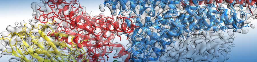
Structure-guided drug design is an integral part of the discovery platform in most pharmaceutical industries. This iterative process can take years and involves many fields of research. The very first step is the determination of the structure of the target of interest. For this, most commonly, methods such as X-ray crystallography (XRD) and nuclear magnetic resonance (NMR) have been used. However, structural information for a large portion of targets of interest for the pharmaceutical industry is still unknown, mainly due to molecule size, complexity, flexibility and difficulties in production and purification. Additionally, structures determined by XRD often represent just one snapshot of one conformation of the target protein. This snapshot does not necessarily represent the most common conformation present at a cellular level and often cannot provide detailed information about flexibility and conformational motions.
As cryo-electron microscopy (cryo-EM) has emerged as a complementary technique to XRD and NMR, it has shown the potential for several advantages over the more traditional methods: access to larger and/or more complex biological systems, analysis of proteins in solutions (vitrified) and, perhaps most importantly, the possibility of characterizing multiple conformational or compositional states from the same sample. This last possibility holds the promise of providing insights into states of the macromolecule theoretically closer to those that are biologically relevant.
In her presentation, Cryo-EM for drug discovery, design, understanding and application, Giovanna Scapin, Sr. Principal Scientist, Computational and Structural Chemistry at Merck & Co., discusses the current state of cryo-EM in the area of small-molecule, structure-guided drug discovery, what it can bring to your research, and what can be done (or expected) in the future to move this technique to the forefront of drug design.