Search
Invitrogen
Aurora A Monoclonal Antibody (35C1)
{{$productOrderCtrl.translations['antibody.pdp.commerceCard.promotion.promotions']}}
{{$productOrderCtrl.translations['antibody.pdp.commerceCard.promotion.viewpromo']}}
{{$productOrderCtrl.translations['antibody.pdp.commerceCard.promotion.promocode']}}: {{promo.promoCode}} {{promo.promoTitle}} {{promo.promoDescription}}. {{$productOrderCtrl.translations['antibody.pdp.commerceCard.promotion.learnmore']}}
Please note: We are reviewing Western blot images included in the antibody testing data in our catalog, including those provided by third parties. Unless expressly labeled or annotated as “raw-unedited”, Western blot images included in the antibody testing data in our catalog may have been edited, optimized or otherwise adjusted for presentation.


产品信息
MA1-80399
应用
建议稀释比
已发表文章
产品规格
宿主/亚型
Mouse
/ IgG2b
分类
Monoclonal
类型
Antibody
克隆号
35C1
抗原
Recombinant Aurora A
偶联物
Unconjugated
Unconjugated
Unconjugated
形式
Liquid
浓度
1 mg/mL
规格
100 µg
保存条件
4°C or -20°C if preferred
运输条件
Wet ice
RRID
产品详细信息
Clone 35C1 specifically recognizes an epitope within the non-catalytic N-terminal domain of Aurora-A, and does not inhibit Aurora-A kinase activity.
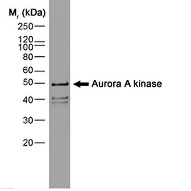
A suggested positive control for immunohistochemical applications is human 293 and mouse llc1 cell lines. In Western blot, this antibody detects a band at ~46kDa in human HeLa and mouse M-ICc12 cell lysates.
靶标信息
AURKA (Aurora A) is a serine/threonine kinase that regulates mitosis by association in vivo with centrosomes. Overexpression of AURKA is observed in many types of cancer. Aurora A plays a role in cell cycle regulation during anaphase and/or telophase, in relation to the function of the centrosome/spindle pole region during chromosome segregation. Aurora A plays a key role during tumor development and progression and is overexpressed in many human cancers including breast, ovarian and colorectal. Aurora A is viewed as a potential target for anticancer drug treatment. Tissue specificity: Highly expressed in testis and weakly in skeletal muscle, thymus and spleen.
仅用于科研。不用于诊断过程。未经明确授权不得转售。
篇参考文献 (0)
您是否在文献中引用过该产品?请点击下方按钮邮件告知我们。
Disclaimer

Clicking the images or links will redirect you to a website hosted by BenchSci that provides third-party scientific content. Neither the content nor the BenchSci technology and processes for selection have been evaluated by us; we are providing them as-is and without warranty of any kind, including for use or application of the Thermo Fisher Scientific products presented.

