Search
Disclaimer

Clicking the images or links will redirect you to a website hosted by BenchSci that provides third-party scientific content. Neither the content nor the BenchSci technology and processes for selection have been evaluated by us; we are providing them as-is and without warranty of any kind, including for use or application of the Thermo Fisher Scientific products presented.
Invitrogen
CD73 Monoclonal Antibody (AD2), NovaFluor™ Red 725, eBioscience™
{{$productOrderCtrl.translations['antibody.pdp.commerceCard.promotion.promotions']}}
{{$productOrderCtrl.translations['antibody.pdp.commerceCard.promotion.viewpromo']}}
{{$productOrderCtrl.translations['antibody.pdp.commerceCard.promotion.promocode']}}: {{promo.promoCode}} {{promo.promoTitle}} {{promo.promoDescription}}. {{$productOrderCtrl.translations['antibody.pdp.commerceCard.promotion.learnmore']}}
")
图: 1 / 2
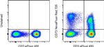
CD73 Antibody (H016T03R05) in Flow
Normal human peripheral blood cells were unstained (left) or stained with CD73 Monoclonal Antibody, NovaFluor Red 725 (Product # H016T03R05) (right). All cells were co-stained with CD3 Monoclonal Antibody, eFluor 450 (Product # 48-0038-82). Total viable cells in the lymphocyte gate were used for analysis, as determined by LIVE/DEAD Blue (Product # L34962). Data was acquired on a 5-laser Cytek Aurora and unmixed with autofluorescence extraction.
 in Flow")
 in Flow")
产品信息
H016T03R05
产品规格
宿主/亚型
Mouse
/ IgG1, kappa
分类
Monoclonal
类型
Antibody
克隆号
AD2
偶联物
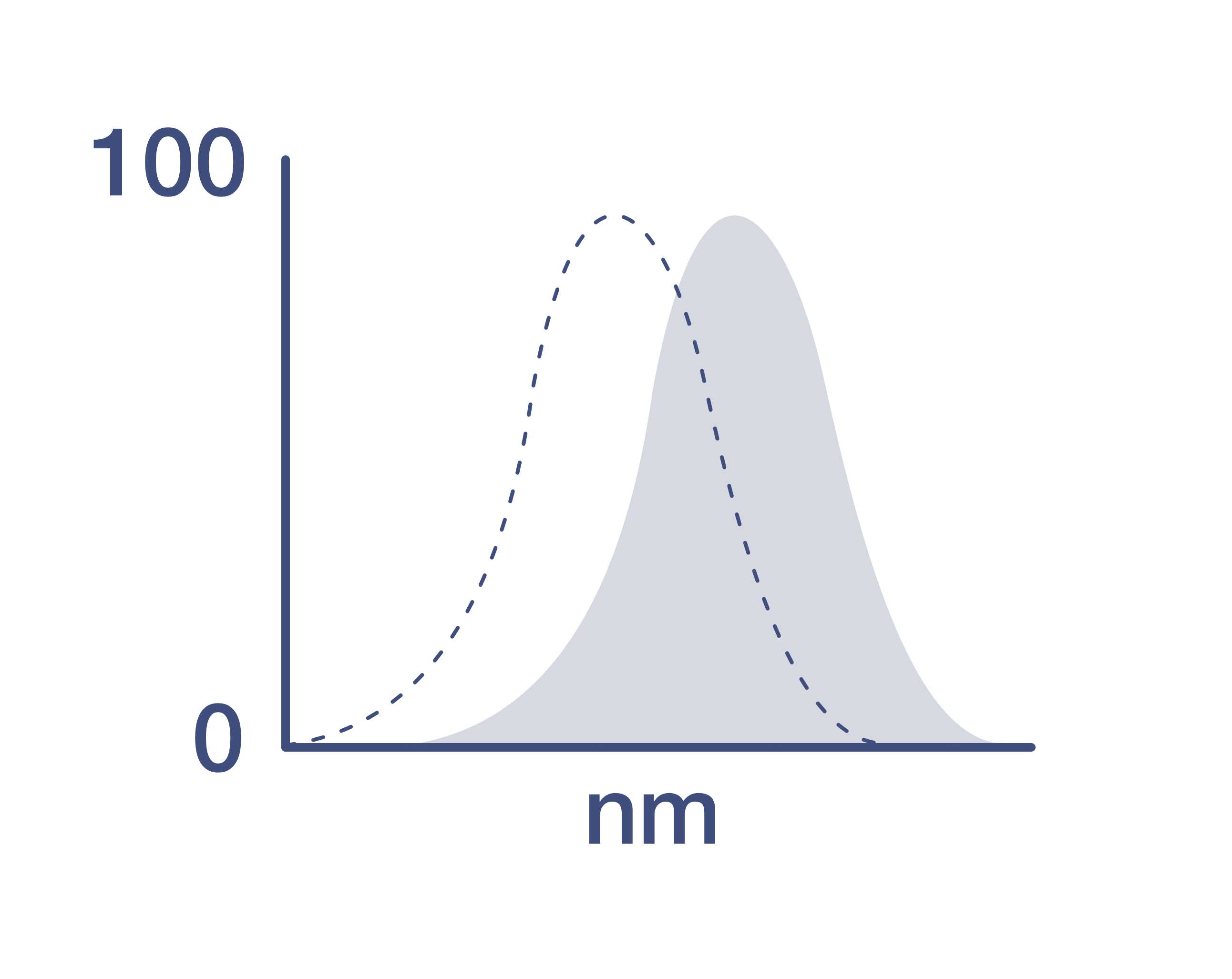
激发/发射光谱
636/727 nm
查看光谱
形式
Liquid
浓度
0.4 µg/Test
保存条件
4°C, store in dark, DO NOT FREEZE!
运输条件
Ambient (domestic); Wet ice (international)
RRID
产品详细信息
Description: This AD2 monoclonal antibody reacts with human CD73, a 5'-ectonucleotidase that converts 5'-adenosine monophosphate to adenosine. CD73 is expressed on the surface of endothelial cells, as well as B and T cells, including some CD4+Foxp3+ regulatory T cells. Adenosine production by these cells has been linked to the inhibition of CD4 T cell effector functions such as proliferation and cytokine secretion.
Applications Reported: This AD2 antibody has been reported for use in flow cytometric analysis.
Applications Tested: This AD2 antibody has been pre-titrated and tested by flow cytometric analysis of normal human peripheral blood cells. This can be used at 5 µL (0.4 µg) per test. A test is defined as the amount (µg) of antibody that will stain a cell sample in a final volume of 100 µL. Cell number should be determined empirically but can range from 10^5 to 10^8 cells/test.
NovaFluor dyes are not compatible with DNA intercalating viability dyes. Do not use viability dyes such as propidium iodide, 7-actinomycin D (7-AAD) and DAPI. Invitrogen LIVE/DEAD Fixable Dead Cell stains are recommended for use with NovaFluor dyes.
Each NovaFluor conjugate or kit is shipped with CellBlox Blocking Buffer. Use this buffer whenever staining with NovaFluor conjugates, including single-color compensation controls using cells. Whenever possible, we recommend adding CellBlox Blocking Buffer to antibody cocktails/master mixes prior to combining with cells. Add 5 µL per sample (regardless of the number of NovaFluors in your panel) to use the antibody cocktail as intended. For single-color controls, use 5 µL of CellBlox Blocking Buffer per 100µL of cell sample containing 10^3 to 10^8 cells.
NovaFluor conjugates are based on Phiton™ technology utilizing novel nucleic acid dye structures that allow for engineered fluorescent signatures with consideration for spillover and spread impacts. Learn more
Excitation: 636 nm; Emission: 727 nm; Laser: 633-640 nm (Red) Laser
靶标信息
CD73, also known as Ecto-5-prime-nucleotidase or 5-prime-ribonucleotide phosphohydrolase, is an enzyme that catalyzes the conversion of purine 5-prime mononucleotides to nucleosides at neutral pH, with AMP as its preferred substrate. It is composed of a dimer of two identical 70 kDa subunits, externally bound to the plasma membrane via a glycosyl phosphatidyl inositol linkage. CD73 serves as a marker of lymphocyte differentiation and is expressed on a subset of lymphocytes, increasing during lymphocyte maturation. It is found on memory CD4 T cells, which resemble uncommitted primed precursor helper cells (Thpp) capable of differentiating into Th1 or Th2 cells, and is also present on regulatory T cells. CD73 deficiency is associated with various immunodeficiency diseases. Other forms of 5-prime nucleotidase exist in the cytoplasm and lysosomes, distinguishable from CD73 by their substrate affinities, requirement for divalent magnesium ions, activation by ATP, and inhibition by inorganic phosphate. The CD73 gene is localized to chromosome 6q14-q21, and defects in this gene can lead to conditions such as calcification of joints and arteries, and intestinal tuberculosis. Additionally, two transcript variants encoding different isoforms of CD73 have been identified.
仅用于科研。不用于诊断过程。未经明确授权不得转售。
How to use the Panel Builder
Watch the video to learn how to use the Invitrogen Flow Cytometry Panel Builder to build your next flow cytometry panel in 5 easy steps.
篇参考文献 (0)
您是否在文献中引用过该产品?请点击下方按钮邮件告知我们。

