Search
Invitrogen
Lamin A/C Monoclonal Antibody (4A7), eBioscience™
{{$productOrderCtrl.translations['antibody.pdp.commerceCard.promotion.promotions']}}
{{$productOrderCtrl.translations['antibody.pdp.commerceCard.promotion.viewpromo']}}
{{$productOrderCtrl.translations['antibody.pdp.commerceCard.promotion.promocode']}}: {{promo.promoCode}} {{promo.promoTitle}} {{promo.promoDescription}}. {{$productOrderCtrl.translations['antibody.pdp.commerceCard.promotion.learnmore']}}
Additional Information:
{{banner.description}}
")
图: 1 / 5
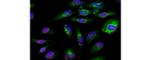
Lamin A/C Antibody (14-9688-80) in ICC/IF
Immunocytochemistry of fixed and permeabilized HeLa cells stained with 10 µg/mL of Anti-Lamin A/C Purified and 10 µg/mL of F(ab')2 Anti-Mouse IgG eFluor® 570 (red), followed by 1 µg/mL of Anti-Pan-Cytokeratin (AE1/AE3) Alexa Fluor® 488 (green).Nuclei are stained with DAPI (blue).

Please note: We are reviewing Western blot images included in the antibody testing data in our catalog, including those provided by third parties. Unless expressly labeled or annotated as “raw-unedited”, Western blot images included in the antibody testing data in our catalog may have been edited, optimized or otherwise adjusted for presentation.
 in ICC/IF")
 in Flow")
 in WB")
 in WB")
")
产品信息
14-9688-80
应用
建议稀释比
已发表文章
产品规格
种属反应
Hamster,
Human,
Mouse,
Rabbit
已发表种属
Mouse
宿主/亚型
Mouse
/ IgG1, kappa
分类
Monoclonal
类型
Antibody
克隆号
4A7
偶联物
Unconjugated
Unconjugated
Unconjugated
形式
Liquid
浓度
0.5 mg/mL
规格
25 µg
纯化类型
Affinity chromatography
保存液
PBS, pH 7.2
内含物
0.09% Sodium Azide
保存条件
4°C
运输条件
Ambient (domestic); Wet ice (international)
RRID
产品详细信息
The monoclonal antibody 4A7 recognizes human, mouse, rabbit and hamster lamin A/C. Lamins are nuclear intermediate filament proteins that provide framework for the nuclear envelope, maintain cell morphology, and protect the nucleus from mechanical, thermal, and oxidative stresses. Lamins also play a role in nuclear assembly, chromatin organization, DNA replication, RNA transcription, cell signaling, and apoptosis. Lamin C is a splice variant of Lamin A and both are A-type lamins. Lamin A and C are crucial for skeletal and cardiac development and function. Defects in A-type lamins result in cardiomyopathy, muscular dystrophy, peripheral neuropathy, lipodystrophy, restrictive dermopathy, and progeroid disorders.
This 4A7 antibody has been tested by immunocytochemistry of methanol-fixed and permeabilized cells and can be used at less than or equal to 10 µg/mL. It is recommended that the antibody be carefully titrated for optimal performance in the assay of interest.
Purity: Greater than 90%, as determined by SDS-PAGE.
Aggregation: Less than 10%, as determined by HPLC.
Filtration: 0.2 µm post-manufacturing filtered.
靶标信息
Lamins are a class of intermediate filament proteins that form a matrix on the inner surface of the nuclear envelope. These proteins are found in many different cell types in three different forms (A, B, and C). Lamins A and C are alternatively spliced versions of the LMNA gene. The LMNA gene has been linked to many disorders of the muscular system, nervous system, and the fat distributions systems including: Emery-Dreifuss muscular dystrophy, Dunnigan-type familial partial lipodystrophy (FPLD), limb-girdle muscular dystrophy (LGMD1B), dilated cardiomyopathy (CMD1A), axonal neuropathy (Charcot-Marie-Tooth disease; CMT2B1), and mandibuloacral dysplasia (MAD).
仅用于科研。不用于诊断过程。未经明确授权不得转售。
生物信息学
蛋白别名: 70 kDa lamin; epididymis secretory sperm binding protein; lamin A protein; Lamin A+C mutant; lamin a-c; lamin A/C; lamin A/C-like 1; Lamin AC; lamin C; lamin C protein; lamin-A/C; mandibuloacral dysplasia type A; mutant 453W; Mutant lamin A/C; Prelamin-A/C; progerin; progerin mutant; renal carcinoma antigen NY-REN-32; RP11-54H19.1; unnamed protein product
基因别名: CDCD1; CDDC; CMD1A; CMT2B1; Dhe; EMD2; FPL; FPLD; FPLD2; HGPS; I79_009616; IDC; Lamin-A/C; LDP1; LFP; LGMD1B; LMN1; LMNA; LMNC; LMNL1; MADA; PRO1
UniProt ID: (Mouse) P48678
Entrez Gene ID: (Human) 4000, (Rabbit) 100343574, (Mouse) 16905, (Chinese hamster) 100757316
nucleus organization
nuclear envelope organization
muscle organ development
establishment of cell polarity
regulation of cell migration
establishment or maintenance of microtubule cytoskeleton polarity
protein localization to nucleus
sterol regulatory element binding protein import into nucleus
ventricular cardiac muscle cell development
negative regulation of release of cytochrome c from mitochondria
positive regulation of cell aging
regulation of protein localization to nucleus
negative regulation of extrinsic apoptotic signaling pathway
double-strand break repair via nonhomologous end joining
nuclear migration
protein localization
negative regulation of cell proliferation
heterochromatin assembly
regulation of telomere maintenance
nuclear pore localization
cellular response to hypoxia
cellular senescence
protein localization to nuclear envelope
negative regulation of cardiac muscle hypertrophy in response to stress
DNA double-strand break attachment to nuclear envelope
Disclaimer

Clicking the images or links will redirect you to a website hosted by BenchSci that provides third-party scientific content. Neither the content nor the BenchSci technology and processes for selection have been evaluated by us; we are providing them as-is and without warranty of any kind, including for use or application of the Thermo Fisher Scientific products presented.

