Search
Invitrogen
Phospho-PLD2 (Tyr169) Polyclonal Antibody
{{$productOrderCtrl.translations['antibody.pdp.commerceCard.promotion.promotions']}}
{{$productOrderCtrl.translations['antibody.pdp.commerceCard.promotion.viewpromo']}}
{{$productOrderCtrl.translations['antibody.pdp.commerceCard.promotion.promocode']}}: {{promo.promoCode}} {{promo.promoTitle}} {{promo.promoDescription}}. {{$productOrderCtrl.translations['antibody.pdp.commerceCard.promotion.learnmore']}}
Additional Information:
{{banner.description}}
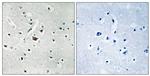
 Antibody in Immunohistochemistry (Paraffin) (IHC (P))")
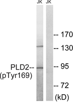
Please note: We are reviewing Western blot images included in the antibody testing data in our catalog, including those provided by third parties. Unless expressly labeled or annotated as “raw-unedited”, Western blot images included in the antibody testing data in our catalog may have been edited, optimized or otherwise adjusted for presentation.
 Antibody (PA5-37689) in IHC (P)")
 Antibody (PA5-37689) in WB")

产品信息
PA5-37689
产品规格
种属反应
Human
宿主/亚型
Rabbit
/ IgG
分类
Polyclonal
类型
Antibody
抗原
Peptide sequence around phosphorylation site of tyrosine 169(E-N-Y(p)-L-N) derived from Human PLD2.
偶联物
Unconjugated
Unconjugated
Unconjugated
形式
Liquid
浓度
1 mg/mL
规格
100 µg
纯化类型
Antigen affinity chromatography
保存液
PBS, pH 7.4, with 50% glycerol
内含物
0.02% sodium azide
保存条件
-20°C
运输条件
Ambient (domestic); Wet ice (international)
RRID
产品详细信息
A suggested positive control for Western blot is Jurkat cells; suggested positive control for IHC is human brain tissue.
靶标信息
PLD2 is a peripheral membrane protein belonging to phospholipase D family with a PH domain, two PLD phosphodiesterase domains and a PX (phox homology) domain that plays a regulatory rather than catalytic role. PLD2 catalyzes the hydrolysis of phosphatidylcholine (PC) to produce phosphatidic acid and choline. PLD2 may function in regulated secretion, signal-induced cytoskeletal regulation and/or endocytosis, transcriptional regulation, and cell cycle control. It is usually stimulated by phosphatidylinositol 4,5-bisphosphate (PIP2) and activated by the ADP-ribosylation factor-1 (ARF-1). PLD2 is activated by agonist stimulation of both tyrosine kinase and G protein-coupled receptors and is also known to interact with EGFR and PIP5K1A. PLD2, localized to plasma membrane caveolae, is ubiquitously expressed in most tissues.
仅用于科研。不用于诊断过程。未经明确授权不得转售。
篇参考文献 (0)
您是否在文献中引用过该产品?请点击下方按钮邮件告知我们。
Disclaimer

Clicking the images or links will redirect you to a website hosted by BenchSci that provides third-party scientific content. Neither the content nor the BenchSci technology and processes for selection have been evaluated by us; we are providing them as-is and without warranty of any kind, including for use or application of the Thermo Fisher Scientific products presented.

