Search
Invitrogen
TCR V alpha 24 J alpha 18 Monoclonal Antibody (6B11), Brilliant Ultra Violet™ 661, eBioscience™
{{$productOrderCtrl.translations['antibody.pdp.commerceCard.promotion.promotions']}}
{{$productOrderCtrl.translations['antibody.pdp.commerceCard.promotion.viewpromo']}}
{{$productOrderCtrl.translations['antibody.pdp.commerceCard.promotion.promocode']}}: {{promo.promoCode}} {{promo.promoTitle}} {{promo.promoDescription}}. {{$productOrderCtrl.translations['antibody.pdp.commerceCard.promotion.learnmore']}}
Additional Information:
{{banner.description}}
")
图: 1 / 1
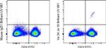
TCR V alpha 24 J alpha 18 Antibody (376-5806-42) in Flow
Normal human peripheral blood cells were stained with CD3 Monoclonal Antibody, FITC (Product # 11-0038-42) and Mouse IgG1 kappa Isotype Control, Brilliant Ultra Violet 661 (Product # 376-4714-81) (left) or TCR V alpha 24 J alpha 18 Monoclonal Antibody, Brilliant Ultra Violet 661 (right). Viable cells in the lymphocyte gate were used for analysis, as determined by Fixable Viability Dye eFluor 780 (Product # ... View More
Please note: We are reviewing Western blot images included in the antibody testing data in our catalog, including those provided by third parties. Unless expressly labeled or annotated as “raw-unedited”, Western blot images included in the antibody testing data in our catalog may have been edited, optimized or otherwise adjusted for presentation.
 in Flow")
产品信息
376-5806-42
产品规格
种属反应
Human
宿主/亚型
Mouse
/ IgG1, kappa
分类
Monoclonal
类型
Antibody
克隆号
6B11
偶联物
Brilliant Ultra Violet™ 661
Brilliant Ultra Violet™ 661
Brilliant Ultra Violet™ 661
激发/发射光谱
349/659 nm
查看光谱
形式
liquid
浓度
5 µL/Test
纯化类型
Affinity chromatography
保存液
PBS, pH 7.2, with BSA
内含物
0.09% sodium azide
保存条件
4°C, store in dark, DO NOT FREEZE!
运输条件
Ambient (domestic); Wet ice (international)
RRID
产品详细信息
Description:
The 6B11 monoclonal antibody reacts with an epitope of the CDR3 formed by the germ-line configuration of the V alpha 24 and J alpha 18 of the TCR alpha locus, which results in the expression of an invariant T-cell receptor (TCR) expressed by invariant NK T cells (iNKT). This TCR is homologous to the murine V alpha 14J alpha 18. This TCR alpha chain pairs with a restricted set of rearranged TCR beta chains, with V beta 11 being the most prominent in humans. NK T cells are restricted by the CD1d molecule and are activated by a CD1d-presented glycolipid ligand, which results in the rapid production of IL-4, IL-13 and IFN gamma. Although iNKT cells have NK-like cytolytic activity, they are considered regulators of immune responses because they rapidly produce large amounts of both Th1 and Th2 cytokines in autoimmune disease, infectious disease, and cancer. The number of iNKT cells present in peripheral blood of normal humans shows considerable variability ranging from 0.01-1% of total peripheral T cells.
Applications Tested:
This 6B11 antibody has been pre-diluted and tested by flow cytometric analysis of normal human peripheral blood cells. This may be used at 5 µL (0.5 µg) per test. A test is defined as the amount (µg) of antibody that will stain a cell sample in a final volume of 100 µL. Cell number should be determined empirically but can range from 10^5 to 10^8 cells/test.
Blocking Buffers
When using two or more Super Bright, Brilliant Violet™, Brilliant Ultra Violet™, or other polymer dye-conjugated antibodies in a staining panel, it is recommended to use Super Bright Complete Staining Buffer (Product # SB-4401) or Brilliant Stain Buffer (Product # 00-4409-75) to minimize any non-specific polymer interactions. Please refer to the datasheet for Super Bright Staining Buffer or Brilliant Stain Buffer for more information.
Light sensitivity
This tandem dye is sensitive to photo-induced oxidation. Please protect this vial and stained samples from light.
Fixation
• Samples can be stored in IC Fixation Buffer (Product # 00-8222) (100 µL of cell sample + 100 µL of IC Fixation Buffer) or 1-step Fix/Lyse Solution (Product # 00-5333) for up to 3 days in the dark at 4°C with minimal impact on brightness and FRET efficiency/compensation.
• Some generalizations regarding fluorophore performance after fixation can be made, but clone specific performance should be determined empirically.
Excitation: 350 nm; Emission: 660 nm; Laser: Ultraviolet Laser.
BRILLIANT ULTRA VIOLET™ is a trademark or registered trademark of Becton, Dickinson and Company or its affiliates, and is used under license. Powered by Sirigen™.
靶标信息
The ability of T cell receptors (TCR) to discriminate foreign from self-peptides presented by major histocompatibility complex (MHC) class II molecules is essential for an effective adaptive immune response. TCR recognition of self-peptides has been linked to autoimmune disease. Mutant self-peptides have been associated with tumors. Engagement of TCRs by a family of bacterial toxins know as superantigens has been responsible for toxic shock syndrome. Autoantibodies to V beta segments of T cell receptors have been isolated from patients with rheumatoid arthritis (RA) and systemic lupus erythematosus (SLE). The autoantibodies block TH1-mediated inflammatory autodestructive reactions and are believed to be a method by which the immune system compensates for disease. Most human T cells express the TCR alpha-beta and either CD4 or CD8 molecule (single positive, SP). A small number of T cells lack both CD4 and CD8 (double negative, DN). Increased percentages of alpha-beta DN T cells have been identified in some autoimmune and immunodeficiency disorders. Gamma-delta T cells are primarily found within the epithelium. They show less TCR diversity and recognize antigens differently than alpha-beta T cells. Subsets of gamma-delta T cells have shown antitumor and immunoregulatory activity.
仅用于科研。不用于诊断过程。未经明确授权不得转售。
How to use the Panel Builder
Watch the video to learn how to use the Invitrogen Flow Cytometry Panel Builder to build your next flow cytometry panel in 5 easy steps.
篇参考文献 (0)
您是否在文献中引用过该产品?请点击下方按钮邮件告知我们。
生物信息学
蛋白别名: Va24; Valpha24
Disclaimer

Clicking the images or links will redirect you to a website hosted by BenchSci that provides third-party scientific content. Neither the content nor the BenchSci technology and processes for selection have been evaluated by us; we are providing them as-is and without warranty of any kind, including for use or application of the Thermo Fisher Scientific products presented.

