Search
Gibco™
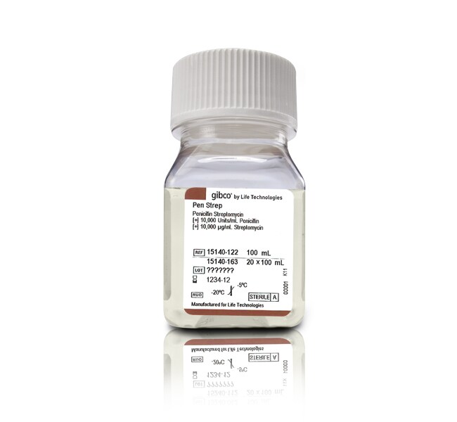
青霉素-链霉素 (10,000 U/mL)
货号:15140122


引用和文献 (34)
Have Questions?
货号 15140122
又称 15140-122
价格(CNY)
344.00
飞享价
Ends: 31-Dec-2026
455.00共减 111.00 (24%)
Each
数量:
100 mL
价格(CNY)
344.00
飞享价
Ends: 31-Dec-2026
455.00共减 111.00 (24%)
Each
更改视图

| 货号 | 数量 |
|---|---|
| 15140122 又称 15140-122 | 100 mL |
| 15140148 又称 15140-148 | 20 mL |
| 15140163 又称 15140-163 | 20 x 100 mL |
产品概述
常见问题解答
规格
文件