Search

Thermo Scientific™
Pierce™ Gaussia 荧光素酶辉光检测试剂盒
在 Gaussia 荧光素酶报告基因存在的情况下获得极其明亮且稳定的生物发光信号(半衰期大于 1 小时)
Have Questions?
更改视图

| 货号 | 数量 |
|---|---|
| 16160 | 100 次反应试剂盒 |
| 16161 | 1000 次反应试剂盒 |
货号 16160
价格(CNY)
1,352.00
Each
数量:
100 次反应试剂盒
价格(CNY)
1,352.00
Each
Thermo Scientific™ Pierce Gaussia 荧光素酶辉光检测试剂盒尤其在 Gaussia-Dura 荧光素酶报告基因存在的情况下提供极其明亮且稳定的生物发光信号(半衰期大于 1 小时)。
Thermo Scientific Pierce Gaussia 荧光素酶辉光检测试剂盒尤其在 Gaussia-Dura 荧光素酶报告基因存在的情况下提供极其明亮且稳定的生物发光信号(半衰期大于 1 小时)。
Gaussia 荧光素酶辉光检测试剂盒的特点:
•灵敏—对 Gaussia 荧光素酶活性进行高度灵敏的检测
• 稳定—更高的信号稳定性
• 简单—不需要使用配备进样器的光度计
• 兼容—与其他 Gaussia 荧光素酶兼容的测定试剂
• 适合自动化—适合用于高通量测定
• 便利—包含一种通用的细胞裂解缓冲液和经过优化的辉光测定试剂
• 安全—允许用户执行非放射性测定
该 Pierce 荧光素酶辉光检测试剂盒包含的试剂用于测定哺乳动物细胞培养基和裂解物中 Gaussia 荧光素酶的活性。当与 Thermo Scientific Gaussia-Dura Luc 载体一起使用时,该试剂盒提供了一个非常灵敏的生物发光报告基因测定系统,用于对荧光素酶活性进行分泌或细胞内检测,提供更高的信号稳定性。荧光素酶反应产生的光输出可与生成的荧光素酶蛋白的量相关,后者又与驱动荧光素酶表达的启动子的活性成正比。该试剂盒可用于检测使用腔肠素作为底物的任何 Gaussia 荧光素酶或衍生物的活性。
包括:
细胞裂解缓冲液、反应缓冲液和底物
需要:
Gaussia 荧光素酶和光度计或其他能够监测发光的仪器(如 Thermo Scientific Luminoskan Ascent 和 Varioskan 闪光微孔板读数仪)。
应用:
• 用于分析顺式调节元素和反式作用因子的启动子研究
• 药物筛选
• siRNA 和 miRNA 筛选
• 研究脱靶效应的多通路测定
• 分泌通路/蛋白定位报告基因测定
• 信号转导通路分析
• RNA 剪接研究
Gaussia 荧光素酶是来自海洋桡脚类动物 Gaussia princeps 的 20kDa 蛋白。生物发光酶在细胞培养基中高度分泌,可对报告基因活性进行活细胞监测。荧光素酶反应产生的光输出可与生成的 Gaussia 荧光素酶蛋白的量相关,并可用于测定驱动 Gaussia 表达的启动子的活性。Gaussia 荧光素酶所产生的信号表现出很强的闪光动力学特征,并且比在类似条件下测定的萤火虫或海肾荧光素酶的信号高得多。
Thermo Scientific Gaussia-Dura Luc 载体中采用的 Gaussia-Dura 基因源自野生型 Gaussia 基因,可提供同样明亮但更稳定的光输出。Gaussia 辉光检测试剂盒试剂可产生辉光型动力学的 Gaussia 荧光素酶反应,并推荐用于不配备进样器的光度计或需要批量处理样品的应用。
仅供科研使用。不可用于诊断程序。
Gaussia 荧光素酶是来自海洋桡脚类动物 Gaussia princeps 的 20kDa 蛋白。生物发光酶在细胞培养基中高度分泌,可对报告基因活性进行活细胞监测。荧光素酶反应产生的光输出可与生成的 Gaussia 荧光素酶蛋白的量相关,并可用于测定驱动 Gaussia 表达的启动子的活性。Gaussia 荧光素酶所产生的信号表现出很强的闪光动力学特征,并且比在类似条件下测定的萤火虫或海肾荧光素酶的信号高得多。
Thermo Scientific Gaussia-Dura Luc 载体中采用的 Gaussia-Dura 基因源自野生型 Gaussia 基因,可提供同样明亮但更稳定的光输出。Gaussia 辉光检测试剂盒试剂可产生辉光型动力学的 Gaussia 荧光素酶反应,并推荐用于不配备进样器的光度计或需要批量处理样品的应用。
参考文献:
- Szent-Gyorgyi, C., et al.(1999).San Jose, CA.Proc.SPIE 3600:4-11.
- Tannous, B. A., et al.(2005).Molecular Therapy 11:435-443.
规格
检测报告基因酶、荧光素酶报告基因检测
适用细胞哺乳动物细胞
检测方法生物发光
适用于(设备)化学发光酶标仪(微孔板)
产品规格384 孔板、96 孔板
标签类型酶标记
产品线Pierce
数量100 次反应试剂盒
底物腔肠素
底物属性化学底物
底物类型荧光素酶底物
足够用于100 次荧光素酶检测
靶标荧光素酶、Gaussia 荧光素酶
技术增强的化学发光法
促进剂EF-1α
类型检测试剂盒
Unit SizeEach
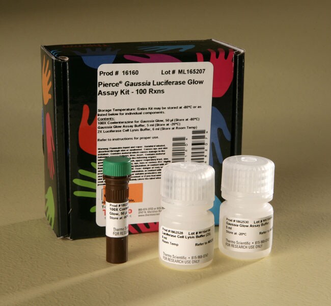
内容与储存
足够用于:100微孔板孔中的荧光素酶测定
• Gaussia 辉光测定缓冲液,5 mL(在 -20°C 下储存)
• 100X 腔肠素,50 μL(在 -80°C 下储存)
• 荧光素酶细胞裂解缓冲液 (2X),6 mL(在室温下储存)
试剂盒可能在 -80°C 下储存或单独储存在指定温度下。
• Gaussia 辉光测定缓冲液,5 mL(在 -20°C 下储存)
• 100X 腔肠素,50 μL(在 -80°C 下储存)
• 荧光素酶细胞裂解缓冲液 (2X),6 mL(在室温下储存)
试剂盒可能在 -80°C 下储存或单独储存在指定温度下。