Search

Applied Biosystems™
MAGnify™ 染色质免疫沉淀系统
MAGnify™ 染色质免疫沉淀系统为使用磁珠捕获技术富集染色质/蛋白复合物和 DNA 回收提供了一种简化、优化测定。分离的 DNA 可以通过基于 PCR 或了解更多信息
| 货号 | 数量 |
|---|---|
| 492024 | 1 个试剂盒 |
货号 492024
价格(CNY)
11,813.00
Each
数量:
1 个试剂盒
价格(CNY)
11,813.00
Each
MAGnify™ 染色质免疫沉淀系统为使用磁珠捕获技术富集染色质/蛋白复合物和 DNA 回收提供了一种简化、优化测定。分离的 DNA 可以通过基于 PCR 或 qPCR 的测定或大量平行 DNA 测序等方法进行下游分析。
ChIP
染色质免疫沉淀 (ChIP) 是研究某些蛋白质与基因组特异性区域相关的强大技术。认为这些序列特异性 DNA 结合蛋白在细胞过程(如 DNA 复制、重组、修复和分离)、染色体稳定性、细胞周期进展和表观遗传沉默中发挥作用。在标准的 ChIP 检测中,通过甲醛处理固定细胞、剪切染色质并通过高特异性抗体进行免疫沉淀。然后研究人员分析 DNA 以确定染色质相关蛋白在体内与染色质结合的基因组区域。该试剂盒使研究人员能够以低于传统 ChIP 工作流程所需的样品量开始测定,从而保留珍贵样品,并且方案可在一天内完成,而传统 ChIP 检测为 2–3 天。该试剂盒可与我们的 ChIP 验证抗体组配合使用,并与 MethylCode™ 和 NCode™产品互补,用于下游表观遗传学研究。
使用 MAGnify™ 系统
使用 MAGnify™ 系统时,您可使用甲醛处理细胞或组织,以在染色质复合物相邻分子间生成蛋白-蛋白和蛋白-DNA 交联。然后裂解细胞,从细胞核中释放染色质并通过超声处理剪切,将平均 DNA 片段大小降低至 200–500 bp 以通过实时荧光定量 PCR (qPCR) 进行分析,或降低至 100–300 bp 以通过大量平行 DNA 测序进行分析。然后免疫沉淀,并使用与 Dynabeads™ 蛋白 A/G 偶联的特异性 ChIP 验证抗体分离目标交联蛋白。通过热处理逆转甲醛交联,并纯化与该蛋白相关的 DNA。DNA 现已准备好用于下游分析,如终点 PCR 或定量 PCR (qPCR)、使用启动子平铺阵列的全基因组分析或下一代测序。在 PCR/qPCR 分析中,引物设计用于跨越所需的目标 DNA 序列,并且数据证明目标特异性蛋白在体内是否与该 DNA 区域相关。
仅供科研使用。不可用于诊断程序。
规格
适用于(应用)染色质免疫沉淀
高通量能力兼容高通量应用
数量1 个试剂盒
样品类型细胞培养物、DNA(基因组)、活细胞
足够用于24 次反应
产品线MAGnify
类型免疫沉淀系统
Unit SizeEach
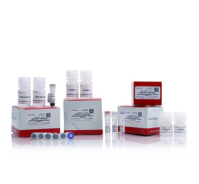
内容与储存
模块 1(湿冰运输,在 4°C 下储存):
• 2 × 1 mL 甘氨酸 (1.25 M)
• 250 µL Dynabeads™蛋白 A/G(切勿冷冻)
•1.4 mL 反相交联缓冲液
• 500 µL DNA 纯化磁珠(切勿冷冻)
• 1.4 mL DNA 纯化缓冲液
• 200 µL 蛋白酶 K (20 mg/mL)
模块 2(湿冰运输,在 4°C 下储存):
• 10 mL IP 缓冲液 1
• 7.5 mL IP 缓冲液 2
• 8 mL DNA 洗涤缓冲液
• 7.2 mL DNA 洗脱缓冲液
模块 3(置于干冰上运输,在 -20°C 下储存):
• 100 µL 蛋白酶抑制剂 (200X)
• 15 µ L 小鼠 IgG (1 µg/µL)
• 15µ L 兔 IgG (1 µg/µL)
模块(置于干冰上运输,在 -20°C 下储存):
• 8 mL 稀释缓冲液
• 3.6 mL 裂解缓冲液